2WNU

Complex between c1q globular heads and heparan sulfate


Experimental Data Snapshot

  • Method: X-RAY DIFFRACTION
  • Resolution: 2.30 Å
  • R-Value Free: 0.256 
  • R-Value Work: 0.198 
  • R-Value Observed: 0.201 

wwPDB Validation   3D Report Full Report


Ligand Structure Quality Assessment 


This is version 2.1 of the entry. See complete history


Literature

Cutting Edge: C1Q Binds Deoxyribose and Heparan Sulfate Through Neighboring Sites of its Recognition Domain.

Garlatti, V.Chouquet, A.Lunardi, T.Vives, R.Paidassi, H.Lortat-Jacob, H.Thielens, N.M.Arlaud, G.J.Gaboriaud, C.

(2010) J Immunol 185: 808

  • DOI: https://doi.org/10.4049/jimmunol.1000184
  • Primary Citation of Related Structures:  
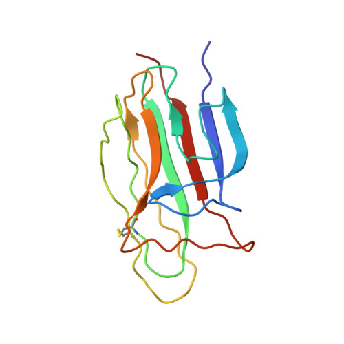
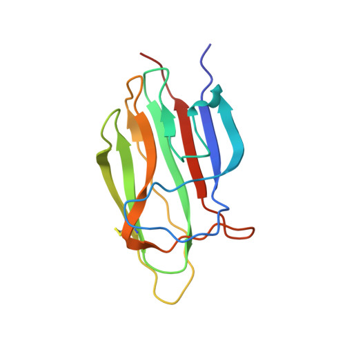
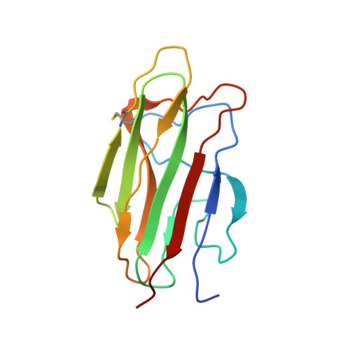
    2WNU, 2WNV

  • PubMed Abstract: 

    C1q, the recognition subunit of the C1 complex of complement, is an archetypal pattern recognition molecule with the striking ability to sense a wide variety of targets, including a number of altered self-motifs. The recognition properties of its globular domain were further deciphered by means of x-ray crystallography using deoxy-D-ribose and heparan sulfate as ligands. Highly specific recognition of deoxy-D-ribose, involving interactions with Arg C98, Arg C111, and Asn C113, was observed at 1.2 A resolution. Heparin-derived tetrasaccharide interacted more loosely through Lys C129, Tyr C155, and Trp C190. These data together with previous findings define a unique binding area exhibiting both polyanion and deoxy-D-ribose recognition properties, located on the inner face of C1q. DNA and heparin compete for C1q binding but are poor C1 activators compared with immune complexes. How the location of this binding area in C1q may regulate the level of C1 activation is discussed.


  • Organizational Affiliation

    Laboratoire de Cristallogenese et Cristallographie des Protéines, Institut de Biologie Structurale Jean-Pierre Ebel, Commissariat à l'Energie Atomique, Centre National de la Recherche Scientifique, Université Joseph Fourier, Grenoble, France.


Macromolecules
Find similar proteins by:  (by identity cutoff)  |  3D Structure
Entity ID: 1
MoleculeChains Sequence LengthOrganismDetailsImage
COMPLEMENT C1Q SUBCOMPONENT SUBUNIT A
A, D
134Homo sapiensMutation(s): 0 
UniProt & NIH Common Fund Data Resources
Find proteins for P02745 (Homo sapiens)
Explore P02745 
Go to UniProtKB:  P02745
PHAROS:  P02745
GTEx:  ENSG00000173372 
Entity Groups  
Sequence Clusters30% Identity50% Identity70% Identity90% Identity95% Identity100% Identity
UniProt GroupP02745
Sequence Annotations
Expand
  • Reference Sequence
Find similar proteins by:  (by identity cutoff)  |  3D Structure
Entity ID: 2
MoleculeChains Sequence LengthOrganismDetailsImage
COMPLEMENT C1Q SUBCOMPONENT SUBUNIT B
B, E
136Homo sapiensMutation(s): 0 
UniProt & NIH Common Fund Data Resources
Find proteins for P02746 (Homo sapiens)
Explore P02746 
Go to UniProtKB:  P02746
PHAROS:  P02746
Entity Groups  
Sequence Clusters30% Identity50% Identity70% Identity90% Identity95% Identity100% Identity
UniProt GroupP02746
Sequence Annotations
Expand
  • Reference Sequence
Find similar proteins by:  (by identity cutoff)  |  3D Structure
Entity ID: 3
MoleculeChains Sequence LengthOrganismDetailsImage
COMPLEMENT C1Q SUBCOMPONENT SUBUNIT C
C, F
131Homo sapiensMutation(s): 0 
UniProt & NIH Common Fund Data Resources
Find proteins for P02747 (Homo sapiens)
Explore P02747 
Go to UniProtKB:  P02747
PHAROS:  P02747
GTEx:  ENSG00000159189 
Entity Groups  
Sequence Clusters30% Identity50% Identity70% Identity90% Identity95% Identity100% Identity
UniProt GroupP02747
Sequence Annotations
Expand
  • Reference Sequence
Oligosaccharides

Help

Entity ID: 4
MoleculeChains Length2D Diagram Glycosylation3D Interactions
2-O-sulfo-beta-L-altropyranuronic acid-(1-4)-2-deoxy-6-O-sulfo-2-(sulfoamino)-alpha-D-glucopyranose
G
2N/A
Glycosylation Resources
GlyTouCan:  G88021JE
GlyCosmos:  G88021JE
Small Molecules
Ligands 1 Unique
IDChains Name / Formula / InChI Key2D Diagram3D Interactions
NAG
Query on NAG

Download Ideal Coordinates CCD File 
H [auth A]2-acetamido-2-deoxy-beta-D-glucopyranose
C8 H15 N O6
OVRNDRQMDRJTHS-FMDGEEDCSA-N
Experimental Data & Validation

Experimental Data

  • Method: X-RAY DIFFRACTION
  • Resolution: 2.30 Å
  • R-Value Free: 0.256 
  • R-Value Work: 0.198 
  • R-Value Observed: 0.201 
  • Space Group: P 1
Unit Cell:
Length ( Å )Angle ( ˚ )
a = 48.09α = 92.86
b = 48.16β = 92.2
c = 87.73γ = 113.64
Software Package:
Software NamePurpose
REFMACrefinement
XDSdata reduction
XSCALEdata scaling
PHASERphasing

Structure Validation

View Full Validation Report


Ligand Structure Quality Assessment 


Entry History 

Deposition Data

Revision History  (Full details and data files)

  • Version 1.0: 2010-05-26
    Type: Initial release
  • Version 1.1: 2011-05-08
    Changes: Version format compliance
  • Version 1.2: 2011-07-13
    Changes: Version format compliance
  • Version 2.0: 2020-07-29
    Type: Remediation
    Reason: Carbohydrate remediation
    Changes: Advisory, Atomic model, Data collection, Derived calculations, Other, Structure summary
  • Version 2.1: 2023-12-13
    Changes: Advisory, Data collection, Database references, Refinement description, Structure summary