2WFO

Crystal structure of Machupo virus envelope glycoprotein GP1


Experimental Data Snapshot

  • Method: X-RAY DIFFRACTION
  • Resolution: 1.73 Å
  • R-Value Free: 0.213 
  • R-Value Work: 0.172 
  • R-Value Observed: 0.174 

wwPDB Validation   3D Report Full Report


Ligand Structure Quality Assessment 


This is version 2.0 of the entry. See complete history


Literature

Unusual Molecular Architecture of the Machupo Virus Attachment Glycoprotein.

Bowden, T.A.Crispin, M.Graham, S.C.Harvey, D.J.Grimes, J.M.Jones, E.Y.Stuart, D.I.

(2009) J Virol 83: 8259

  • DOI: https://doi.org/10.1128/JVI.00761-09
  • Primary Citation of Related Structures:  
    2WFO

  • PubMed Abstract: 

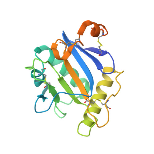
    New World arenaviruses, which cause severe hemorrhagic fever, rely upon their envelope glycoproteins for attachment and fusion into their host cell. Here we present the crystal structure of the Machupo virus GP1 attachment glycoprotein, which is responsible for high-affinity binding at the cell surface to the transferrin receptor. This first structure of an arenavirus glycoprotein shows that GP1 consists of a novel alpha/beta fold. This provides a blueprint of the New World arenavirus attachment glycoproteins and reveals a new architecture of viral attachment, using a protein fold of unknown origins.


  • Organizational Affiliation

    University of Oxford, United Kingdom.


Macromolecules
Find similar proteins by:  (by identity cutoff)  |  3D Structure
Entity ID: 1
MoleculeChains Sequence LengthOrganismDetailsImage
GLYCOPROTEIN 1182Mammarenavirus machupoenseMutation(s): 0 
UniProt
Find proteins for Q6PXS3 (Machupo virus)
Explore Q6PXS3 
Go to UniProtKB:  Q6PXS3
Entity Groups  
Sequence Clusters30% Identity50% Identity70% Identity90% Identity95% Identity100% Identity
UniProt GroupQ6PXS3
Sequence Annotations
Expand
  • Reference Sequence
Oligosaccharides

Help

Entity ID: 2
MoleculeChains Length2D Diagram Glycosylation3D Interactions
2-acetamido-2-deoxy-beta-D-glucopyranose-(1-4)-2-acetamido-2-deoxy-beta-D-glucopyranose
B
2N-Glycosylation
Glycosylation Resources
GlyTouCan:  G42666HT
GlyCosmos:  G42666HT
GlyGen:  G42666HT
Experimental Data & Validation

Experimental Data

  • Method: X-RAY DIFFRACTION
  • Resolution: 1.73 Å
  • R-Value Free: 0.213 
  • R-Value Work: 0.172 
  • R-Value Observed: 0.174 
  • Space Group: P 21 21 21
Unit Cell:
Length ( Å )Angle ( ˚ )
a = 51.523α = 90
b = 55.07β = 90
c = 61.327γ = 90
Software Package:
Software NamePurpose
REFMACrefinement
HKL-2000data reduction
SCALEPACKdata scaling
SHARPphasing

Structure Validation

View Full Validation Report


Ligand Structure Quality Assessment 


Entry History 

Deposition Data

Revision History  (Full details and data files)

  • Version 1.0: 2009-06-16
    Type: Initial release
  • Version 1.1: 2011-05-08
    Changes: Version format compliance
  • Version 1.2: 2011-07-13
    Changes: Version format compliance
  • Version 1.3: 2018-02-28
    Changes: Source and taxonomy
  • Version 2.0: 2020-07-29
    Type: Remediation
    Reason: Carbohydrate remediation
    Changes: Atomic model, Data collection, Derived calculations, Other, Structure summary