2WDA

The X-ray structure of the Streptomyces coelicolor A3 Chondroitin AC Lyase in Complex with Chondroitin sulphate


Experimental Data Snapshot

  • Method: X-RAY DIFFRACTION
  • Resolution: 2.30 Å
  • R-Value Free: 0.193 
  • R-Value Work: 0.149 
  • R-Value Observed: 0.151 

wwPDB Validation   3D Report Full Report


This is version 2.1 of the entry. See complete history


Literature

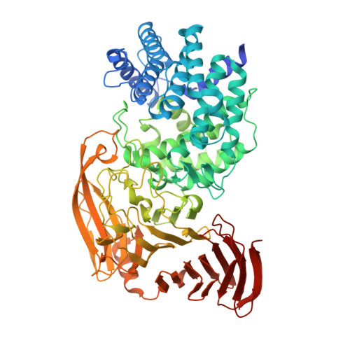
Crystal Structures of a Family 8 Polysaccharide Lyase Reveal Open and Highly Occluded Substrate-Binding Cleft Conformations.

Elmabrouk, Z.H.Vincent, F.Zhang, M.Smith, N.L.Turkenburg, J.P.Charnock, S.J.Black, G.W.Taylor, E.J.

(2011) Proteins 79: 965

  • DOI: https://doi.org/10.1002/prot.22938
  • Primary Citation of Related Structures:  
    2WCO, 2WDA, 2X03

  • PubMed Abstract: 

    Bacterial enzymatic degradation of glycosaminoglycans such as hyaluronan and chondroitin is facilitated by polysaccharide lyases. Family 8 polysaccharide lyase (PL8) enzymes contain at least two domains: one predominantly composed of α-helices, the α-domain, and another predominantly composed of β-sheets, the β-domain. Simulation flexibility analyses indicate that processive exolytic cleavage of hyaluronan, by PL8 hyaluronate lyases, is likely to involve an interdomain shift, resulting in the opening/closing of the substrate-binding cleft between the α- and β-domains, facilitating substrate translocation. Here, the Streptomyces coelicolor A3(2) PL8 enzyme was recombinantly expressed in and purified from Escherichia coli and biochemically characterized as a hyaluronate lyase. By using X-ray crystallography its structure was solved in complex with hyaluronan and chondroitin disaccharides. These findings show key catalytic interactions made by the different substrates, and on comparison with all other PL8 structures reveals that the substrate-binding cleft of the S. coelicolor enzyme is highly occluded. A third structure of the enzyme, harboring a mutation of the catalytic tyrosine, created via site-directed mutagenesis, interestingly revealed an interdomain shift that resulted in the opening of the substrate-binding cleft. These results add further support to the proposed processive mechanism of action of PL8 hyaluronate lyases and may indicate that the mechanism of action is likely to be universally used by PL8 hyaluronate lyases.


  • Organizational Affiliation

    Department of Biomedical Sciences, School of Life Sciences, Northumbria University, Newcastle upon Tyne NE1 8ST, United Kingdom.


Macromolecules
Find similar proteins by:  (by identity cutoff)  |  3D Structure
Entity ID: 1
MoleculeChains Sequence LengthOrganismDetailsImage
PUTATIVE SECRETED LYASE765Streptomyces violaceoruberMutation(s): 0 
EC: 4.2.2.1
UniProt
Find proteins for O86516 (Streptomyces coelicolor (strain ATCC BAA-471 / A3(2) / M145))
Explore O86516 
Go to UniProtKB:  O86516
Entity Groups  
Sequence Clusters30% Identity50% Identity70% Identity90% Identity95% Identity100% Identity
UniProt GroupO86516
Sequence Annotations
Expand
  • Reference Sequence
Oligosaccharides

Help

Entity ID: 2
MoleculeChains Length2D Diagram Glycosylation3D Interactions
4-deoxy-alpha-L-threo-hex-4-enopyranuronic acid-(1-3)-2-acetamido-2-deoxy-4-O-sulfo-beta-D-galactopyranose
B
2N/A
Glycosylation Resources
GlyTouCan:  G76516UK
GlyCosmos:  G76516UK
Small Molecules
Ligands 4 Unique
IDChains Name / Formula / InChI Key2D Diagram3D Interactions
PEG
Query on PEG

Download Ideal Coordinates CCD File 
K [auth A]
L [auth A]
M [auth A]
N [auth A]
O [auth A]
K [auth A],
L [auth A],
M [auth A],
N [auth A],
O [auth A],
P [auth A],
Q [auth A],
R [auth A],
S [auth A]
DI(HYDROXYETHYL)ETHER
C4 H10 O3
MTHSVFCYNBDYFN-UHFFFAOYSA-N
GOL
Query on GOL

Download Ideal Coordinates CCD File 
H [auth A],
I [auth A],
J [auth A]
GLYCEROL
C3 H8 O3
PEDCQBHIVMGVHV-UHFFFAOYSA-N
FMT
Query on FMT

Download Ideal Coordinates CCD File 
C [auth A],
D [auth A],
E [auth A],
F [auth A],
G [auth A]
FORMIC ACID
C H2 O2
BDAGIHXWWSANSR-UHFFFAOYSA-N
MG
Query on MG

Download Ideal Coordinates CCD File 
T [auth A]MAGNESIUM ION
Mg
JLVVSXFLKOJNIY-UHFFFAOYSA-N
Experimental Data & Validation

Experimental Data

  • Method: X-RAY DIFFRACTION
  • Resolution: 2.30 Å
  • R-Value Free: 0.193 
  • R-Value Work: 0.149 
  • R-Value Observed: 0.151 
  • Space Group: P 43 21 2
Unit Cell:
Length ( Å )Angle ( ˚ )
a = 140.508α = 90
b = 140.508β = 90
c = 100.764γ = 90
Software Package:
Software NamePurpose
REFMACrefinement
MOSFLMdata reduction
SCALAdata scaling
PHASERphasing

Structure Validation

View Full Validation Report


Entry History 

Deposition Data

Revision History  (Full details and data files)

  • Version 1.0: 2010-04-21
    Type: Initial release
  • Version 1.1: 2011-05-08
    Changes: Version format compliance
  • Version 1.2: 2011-07-13
    Changes: Version format compliance
  • Version 2.0: 2020-07-29
    Type: Remediation
    Reason: Carbohydrate remediation
    Changes: Atomic model, Data collection, Derived calculations, Non-polymer description, Other, Structure summary
  • Version 2.1: 2023-12-13
    Changes: Data collection, Database references, Refinement description, Structure summary