2VM8

Human CRMP-2 crystallised in the presence of Mg


Experimental Data Snapshot

  • Method: X-RAY DIFFRACTION
  • Resolution: 1.90 Å
  • R-Value Free: 0.325 
  • R-Value Work: 0.256 
  • R-Value Observed: 0.259 

wwPDB Validation   3D Report Full Report


This is version 1.4 of the entry. See complete history


Literature

Crystal and Solution Structure, Stability and Post- Translational Modifications of Collapsin Response Mediator Protein 2.

Majava, V.Loytynoja, N.Chen, W.Q.Lubec, G.Kursula, P.

(2008) FEBS J 275: 4583

  • DOI: https://doi.org/10.1111/j.1742-4658.2008.06601.x
  • Primary Citation of Related Structures:  
    2VM8

  • PubMed Abstract: 

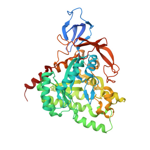
    The collapsin response mediator protein 2 (CRMP-2) is a central molecule regulating axonal growth cone guidance. It interacts with the cytoskeleton and mediates signals related to myelin-induced axonal growth inhibition. CRMP-2 has also been characterized as a constituent of neurofibrillary tangles in Alzheimer's disease. CD spectroscopy and thermal stability assays using the Thermofluor method indicated that Ca2+ and Mg2+ affect the stability of CRMP-2 and prevent the formation of beta-aggregates upon heating. Gel filtration showed that the presence of Ca2+ or Mg2+ promoted the formation of CRMP-2 homotetramers, and this was further proven by small-angle X-ray scattering experiments, where a 3D solution structure for CRMP-2 was obtained. Previously, we described a crystal structure of human CRMP-2 complexed with calcium. In the present study, we determined the structure of CRMP-2 in the absence of calcium at 1.9 A resolution. When Ca2+ was omitted, crystals could only be grown in the presence of Mg2+ ions. By a proteomic approach, we further identified a number of post-translational modifications in CRMP-2 from rat brain hippocampus and mapped them onto the crystal structure.


  • Organizational Affiliation

    Department of Biochemistry, University of Oulu, Finland.


Macromolecules
Find similar proteins by:  (by identity cutoff)  |  3D Structure
Entity ID: 1
MoleculeChains Sequence LengthOrganismDetailsImage
DIHYDROPYRIMIDINASE-RELATED PROTEIN 2
A, B, C, D
501Homo sapiensMutation(s): 0 
UniProt & NIH Common Fund Data Resources
Find proteins for Q16555 (Homo sapiens)
Explore Q16555 
Go to UniProtKB:  Q16555
PHAROS:  Q16555
GTEx:  ENSG00000092964 
Entity Groups  
Sequence Clusters30% Identity50% Identity70% Identity90% Identity95% Identity100% Identity
UniProt GroupQ16555
Sequence Annotations
Expand
  • Reference Sequence
Experimental Data & Validation

Experimental Data

  • Method: X-RAY DIFFRACTION
  • Resolution: 1.90 Å
  • R-Value Free: 0.325 
  • R-Value Work: 0.256 
  • R-Value Observed: 0.259 
  • Space Group: P 21 21 21
Unit Cell:
Length ( Å )Angle ( ˚ )
a = 86.51α = 90
b = 126.24β = 90
c = 209.75γ = 90
Software Package:
Software NamePurpose
REFMACrefinement
XDSdata reduction
XSCALEdata scaling
MOLREPphasing

Structure Validation

View Full Validation Report


Entry History 

Deposition Data

  • Released Date: 2008-08-26 
  • Deposition Author(s): Kursula, P.

Revision History  (Full details and data files)

  • Version 1.0: 2008-08-26
    Type: Initial release
  • Version 1.1: 2011-05-08
    Changes: Version format compliance
  • Version 1.2: 2011-07-13
    Changes: Version format compliance
  • Version 1.3: 2018-01-17
    Changes: Data collection
  • Version 1.4: 2023-12-13
    Changes: Data collection, Database references, Derived calculations, Other, Refinement description