2VKX

Human NCAM, FN3 domains 1 and 2, M610R mutant


Experimental Data Snapshot

  • Method: X-RAY DIFFRACTION
  • Resolution: 2.70 Å
  • R-Value Free: 0.268 
  • R-Value Work: 0.223 
  • R-Value Observed: 0.223 

wwPDB Validation   3D Report Full Report


This is version 1.2 of the entry. See complete history


Literature

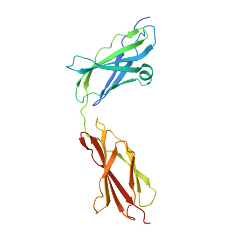
Structure of the Tandem Fibronectin Type 3 Domains of Neural Cell Adhesion Molecule

Carafoli, F.Saffell, J.L.Hohenester, E.

(2008) J Mol Biol 377: 524

  • DOI: https://doi.org/10.1016/j.jmb.2008.01.030
  • Primary Citation of Related Structures:  
    2VKW, 2VKX

  • PubMed Abstract: 

    Activation of the fibroblast growth factor receptor (FGFR) by neural cell adhesion molecule (NCAM) is essential for NCAM-mediated neurite outgrowth. Previous peptide studies have identified two regions in the fibronectin type 3 (FN3)-like domains of NCAM as being important for these activities. Here we report the crystal structure of the NCAM FN3 domain tandem, which reveals an acutely bent domain arrangement. Mutation of a non-conserved surface residue (M610R) led to a second crystal form showing a substantially different conformation. Thus, the FN3 domain linker is highly flexible, suggesting that it corresponds to the hinge seen in electron micrographs of NCAM. The two putative FGFR1-binding segments, one in each NCAM FN3 domain, are situated close to the domain interface. They form a contiguous patch in the more severely bent conformation but become separated upon straightening of the FN3 tandem, suggesting that conformational changes within NCAM may modulate FGFR1 activation. Surface plasmon resonance experiments demonstrated only a very weak interaction between the NCAM FN3 tandem and soluble FGFR1 proteins expressed in mammalian cells (dissociation constant >100 muM). Thus, the NCAM-FGFR1 interaction at the cell surface is likely to depend upon avidity effects due to receptor clustering.


  • Organizational Affiliation

    Department of Life Sciences, Biophysics Section, Blackett Laboratory, Imperial College London, London SW7 2AZ, UK.


Macromolecules
Find similar proteins by:  (by identity cutoff)  |  3D Structure
Entity ID: 1
MoleculeChains Sequence LengthOrganismDetailsImage
NEURAL CELL ADHESION MOLECULE
A, B, C, D, E
A, B, C, D, E, F
209Homo sapiensMutation(s): 1 
UniProt & NIH Common Fund Data Resources
Find proteins for P13591 (Homo sapiens)
Explore P13591 
Go to UniProtKB:  P13591
PHAROS:  P13591
GTEx:  ENSG00000149294 
Entity Groups  
Sequence Clusters30% Identity50% Identity70% Identity90% Identity95% Identity100% Identity
UniProt GroupP13591
Sequence Annotations
Expand
  • Reference Sequence
Small Molecules
Ligands 1 Unique
IDChains Name / Formula / InChI Key2D Diagram3D Interactions
SO4
Query on SO4

Download Ideal Coordinates CCD File 
G [auth A]
H [auth A]
I [auth C]
J [auth C]
K [auth D]
G [auth A],
H [auth A],
I [auth C],
J [auth C],
K [auth D],
L [auth E],
M [auth F],
N [auth F],
O [auth F],
P [auth F],
Q [auth F]
SULFATE ION
O4 S
QAOWNCQODCNURD-UHFFFAOYSA-L
Experimental Data & Validation

Experimental Data

  • Method: X-RAY DIFFRACTION
  • Resolution: 2.70 Å
  • R-Value Free: 0.268 
  • R-Value Work: 0.223 
  • R-Value Observed: 0.223 
  • Space Group: P 21 21 21
Unit Cell:
Length ( Å )Angle ( ˚ )
a = 92.419α = 90
b = 107.573β = 90
c = 161.124γ = 90
Software Package:
Software NamePurpose
CNSrefinement
MOSFLMdata reduction
SCALAdata scaling
PHASERphasing

Structure Validation

View Full Validation Report


Entry History 

Deposition Data

Revision History  (Full details and data files)

  • Version 1.0: 2008-02-26
    Type: Initial release
  • Version 1.1: 2011-05-08
    Changes: Version format compliance
  • Version 1.2: 2011-07-13
    Changes: Version format compliance