2V70

Third LRR domain of human Slit2


Experimental Data Snapshot

  • Method: X-RAY DIFFRACTION
  • Resolution: 3.01 Å
  • R-Value Free: 0.271 
  • R-Value Work: 0.222 
  • R-Value Observed: 0.225 

wwPDB Validation   3D Report Full Report


Ligand Structure Quality Assessment 


This is version 1.3 of the entry. See complete history


Literature

Production of Slit2 Lrr Domains in Mammalian Cells for Structural Studies and the Structure of Human Slit2 Domain 3.

Morlot, C.Hemrika, W.Romijn, R.A.Gros, P.Cusack, S.Mccarthy, A.A.

(2007) Acta Crystallogr D Biol Crystallogr 63: 961

  • DOI: https://doi.org/10.1107/S0907444907035470
  • Primary Citation of Related Structures:  
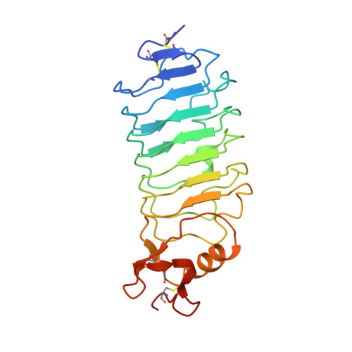
    2V70

  • PubMed Abstract: 

    Slit2 and Roundabout 1 (Robo1) provide a key ligand-receptor interaction for the navigation of commissural neurons during the development of the central nervous system. Slit2 is a large multidomain protein containing an unusual domain organization of four tandem leucine-rich repeat (LRR) domains at its N-terminus. These domains are well known to mediate protein-protein interactions; indeed, the Robo1-binding region has been mapped to the concave face of the second LRR domain. It has also been shown that the fourth LRR domain may mediate Slit dimerization and that both the first and second domains can bind heparin. Thus, while roles have been ascribed for three of the LRR domains, there is still no known role for the third domain. Each of the four LRR domains from human Slit2 have now been successfully expressed in milligram quantities using expression in mammalian cells. Here, the crystallization of the second and third LRR domains and the structure of the third LRR domain are presented. This is the first structure of an LRR domain from human Slit2, which has an extra repeat compared with the Drosophila homologue. It is proposed that a highly conserved patch of surface residues on the concave face may mediate any protein-protein interactions involving this LRR domain, a result that will be useful in guiding further studies on Slit2.


  • Organizational Affiliation

    European Molecular Biology Laboratory, 6 Rue Jules Horowitz, BP 181, 38042 Grenoble, France.


Macromolecules
Find similar proteins by:  (by identity cutoff)  |  3D Structure
Entity ID: 1
MoleculeChains Sequence LengthOrganismDetailsImage
SLIT HOMOLOG 2 PROTEIN N-PRODUCT
A, B, C, D
220Homo sapiensMutation(s): 0 
UniProt & NIH Common Fund Data Resources
Find proteins for O94813 (Homo sapiens)
Explore O94813 
Go to UniProtKB:  O94813
PHAROS:  O94813
GTEx:  ENSG00000145147 
Entity Groups  
Sequence Clusters30% Identity50% Identity70% Identity90% Identity95% Identity100% Identity
UniProt GroupO94813
Sequence Annotations
Expand
  • Reference Sequence
Experimental Data & Validation

Experimental Data

  • Method: X-RAY DIFFRACTION
  • Resolution: 3.01 Å
  • R-Value Free: 0.271 
  • R-Value Work: 0.222 
  • R-Value Observed: 0.225 
  • Space Group: P 1 21 1
Unit Cell:
Length ( Å )Angle ( ˚ )
a = 63.65α = 90
b = 96.86β = 95.32
c = 87.15γ = 90
Software Package:
Software NamePurpose
REFMACrefinement
XDSdata reduction
XSCALEdata scaling
PHASERphasing

Structure Validation

View Full Validation Report


Ligand Structure Quality Assessment 


Entry History 

Deposition Data

Revision History  (Full details and data files)

  • Version 1.0: 2007-08-28
    Type: Initial release
  • Version 1.1: 2011-07-13
    Changes: Advisory, Version format compliance
  • Version 1.2: 2020-07-29
    Type: Remediation
    Reason: Carbohydrate remediation
    Changes: Data collection, Derived calculations, Other, Structure summary
  • Version 1.3: 2023-12-13
    Changes: Data collection, Database references, Refinement description, Structure summary