2V5Y

Crystal structure of the receptor protein tyrosine phosphatase mu ectodomain


Experimental Data Snapshot

  • Method: X-RAY DIFFRACTION
  • Resolution: 3.10 Å
  • R-Value Free: 0.320 
  • R-Value Work: 0.242 
  • R-Value Observed: 0.246 

wwPDB Validation   3D Report Full Report


Ligand Structure Quality Assessment 


This is version 1.3 of the entry. See complete history


Literature

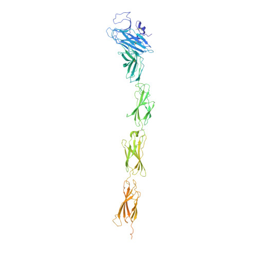
Structure of a Tyrosine Phosphatase Adhesive Interaction Reveals a Spacer-Clamp Mechanism.

Aricescu, A.R.Siebold, C.Choudhuri, K.Chang, V.T.Lu, W.Davis, S.J.Van Der Merwe, P.A.Jones, E.Y.

(2007) Science 317: 1217

  • DOI: https://doi.org/10.1126/science.1144646
  • Primary Citation of Related Structures:  
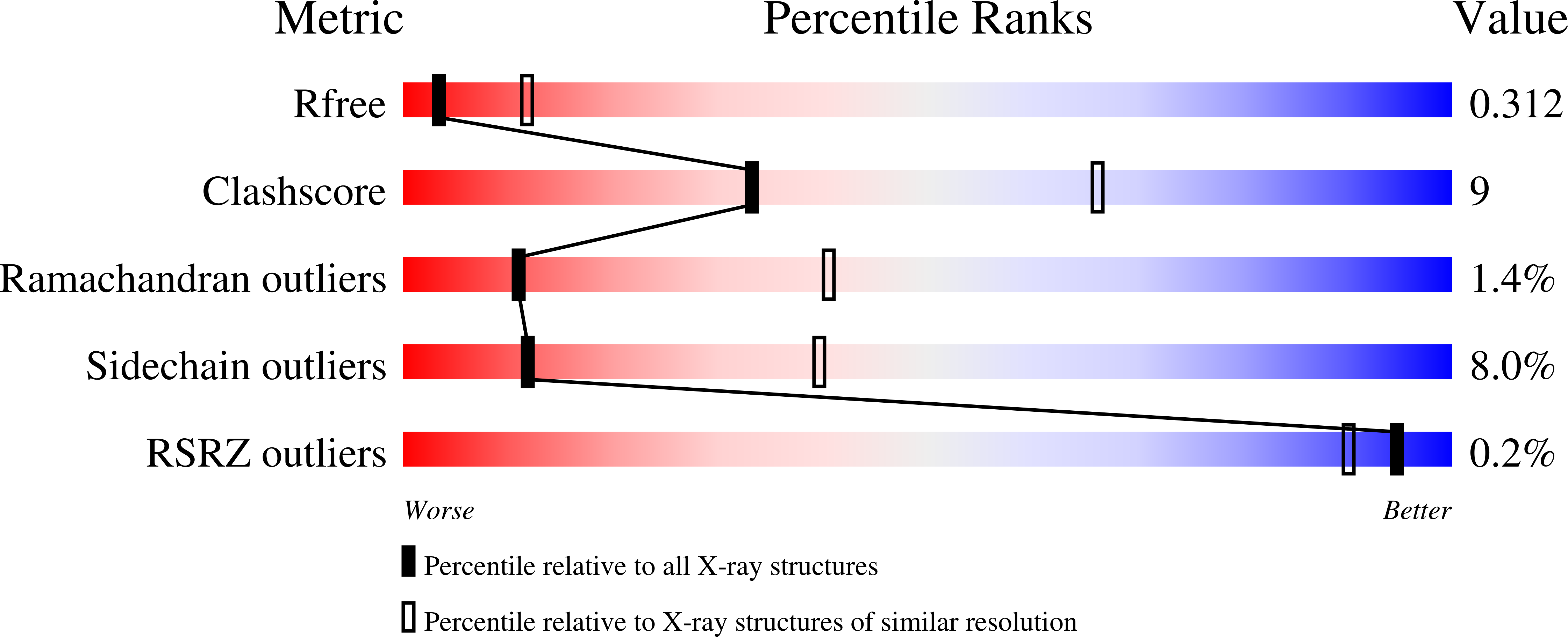
    2V5Y

  • PubMed Abstract: 

    Cell-cell contacts are fundamental to multicellular organisms and are subject to exquisite levels of control. Human RPTPmu is a type IIB receptor protein tyrosine phosphatase that both forms an adhesive contact itself and is involved in regulating adhesion by dephosphorylating components of cadherin-catenin complexes. Here we describe a 3.1 angstrom crystal structure of the RPTPmu ectodomain that forms a homophilic trans (antiparallel) dimer with an extended and rigid architecture, matching the dimensions of adherens junctions. Cell surface expression of deletion constructs induces intercellular spacings that correlate with the ectodomain length. These data suggest that the RPTPmu ectodomain acts as a distance gauge and plays a key regulatory function, locking the phosphatase to its appropriate functional location.


  • Organizational Affiliation

    Cancer Research UK Receptor Structure Research Group, University of Oxford, Henry Wellcome Building of Genomic Medicine, Division of Structural Biology, Roosevelt Drive, Oxford OX3 7BN, UK.


Macromolecules
Find similar proteins by:  (by identity cutoff)  |  3D Structure
Entity ID: 1
MoleculeChains Sequence LengthOrganismDetailsImage
RECEPTOR-TYPE TYROSINE-PROTEIN PHOSPHATASE MU731Homo sapiensMutation(s): 0 
EC: 3.1.3.48
UniProt & NIH Common Fund Data Resources
Find proteins for P28827 (Homo sapiens)
Explore P28827 
Go to UniProtKB:  P28827
PHAROS:  P28827
GTEx:  ENSG00000173482 
Entity Groups  
Sequence Clusters30% Identity50% Identity70% Identity90% Identity95% Identity100% Identity
UniProt GroupP28827
Sequence Annotations
Expand
  • Reference Sequence
Small Molecules
Ligands 2 Unique
IDChains Name / Formula / InChI Key2D Diagram3D Interactions
NAG
Query on NAG

Download Ideal Coordinates CCD File 
B [auth A]
C [auth A]
D [auth A]
E [auth A]
F [auth A]
B [auth A],
C [auth A],
D [auth A],
E [auth A],
F [auth A],
G [auth A],
H [auth A]
2-acetamido-2-deoxy-beta-D-glucopyranose
C8 H15 N O6
OVRNDRQMDRJTHS-FMDGEEDCSA-N
NA
Query on NA

Download Ideal Coordinates CCD File 
I [auth A],
J [auth A]
SODIUM ION
Na
FKNQFGJONOIPTF-UHFFFAOYSA-N
Experimental Data & Validation

Experimental Data

  • Method: X-RAY DIFFRACTION
  • Resolution: 3.10 Å
  • R-Value Free: 0.320 
  • R-Value Work: 0.242 
  • R-Value Observed: 0.246 
  • Space Group: C 1 2 1
Unit Cell:
Length ( Å )Angle ( ˚ )
a = 167.336α = 90
b = 69.325β = 113.52
c = 97.487γ = 90
Software Package:
Software NamePurpose
HKL-2000data reduction
SCALEPACKdata scaling
MOLREPphasing
PHASERphasing
REFMACrefinement

Structure Validation

View Full Validation Report


Ligand Structure Quality Assessment 


Entry History 

Deposition Data

Revision History  (Full details and data files)

  • Version 1.0: 2007-09-11
    Type: Initial release
  • Version 1.1: 2011-07-13
    Changes: Advisory, Version format compliance
  • Version 1.2: 2020-07-29
    Type: Remediation
    Reason: Carbohydrate remediation
    Changes: Data collection, Derived calculations, Other, Structure summary
  • Version 1.3: 2023-12-13
    Changes: Data collection, Database references, Refinement description, Structure summary