2V5T

Crystal structure of NCAM2 Ig2-3


Experimental Data Snapshot

  • Method: X-RAY DIFFRACTION
  • Resolution: 2.00 Å
  • R-Value Free: 0.234 
  • R-Value Work: 0.181 

wwPDB Validation   3D Report Full Report


Ligand Structure Quality Assessment 


This is version 1.3 of the entry. See complete history


Literature

Structural Model and Trans-Interaction of the Entire Ectodomain of the Olfactory Cell Adhesion Molecule.

Kulahin, N.Kristensen, O.Rasmussen, K.K.Olsen, L.Rydberg, P.Vestergaard, B.Kastrup, J.S.Berezin, V.Bock, E.Walmod, P.S.Gajhede, M.

(2011) Structure 19: 203

  • DOI: https://doi.org/10.1016/j.str.2010.12.014
  • Primary Citation of Related Structures:  
    2JLL, 2V5T, 2WIM, 2XY1, 2XY2, 2XYC

  • PubMed Abstract: 

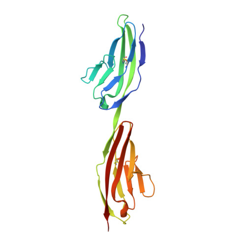
    The ectodomain of olfactory cell adhesion molecule (OCAM/NCAM2/RNCAM) consists of five immunoglobulin (Ig) domains (IgI-V), followed by two fibronectin-type 3 (Fn3) domains (Fn3I-II). A complete structural model of the entire ectodomain of human OCAM has been assembled from crystal structures of six recombinant proteins corresponding to different regions of the ectodomain. The model is the longest experimentally based composite structural model of an entire IgCAM ectodomain. It displays an essentially linear arrangement of IgI-V, followed by bends between IgV and Fn3I and between Fn3I and Fn3II. Proteins containing IgI-IgII domains formed stable homodimers in solution and in crystals. Dimerization could be disrupted in vitro by mutations in the dimer interface region. In conjunction with the bent ectodomain conformation, which can position IgI-V parallel with the cell surface, the IgI-IgII dimerization enables OCAM-mediated trans-interactions with an intercellular distance of about 20 nm, which is consistent with that observed in synapses.


  • Organizational Affiliation

    Protein Laboratory, Department of Neuroscience and Pharmacology, Faculty of Health Sciences, University of Copenhagen, DK-2200 Copenhagen N, Denmark.


Macromolecules
Find similar proteins by:  (by identity cutoff)  |  3D Structure
Entity ID: 1
MoleculeChains Sequence LengthOrganismDetailsImage
NEURAL CELL ADHESION MOLECULE 2189Homo sapiensMutation(s): 0 
UniProt & NIH Common Fund Data Resources
Find proteins for O15394 (Homo sapiens)
Explore O15394 
Go to UniProtKB:  O15394
PHAROS:  O15394
GTEx:  ENSG00000154654 
Entity Groups  
Sequence Clusters30% Identity50% Identity70% Identity90% Identity95% Identity100% Identity
UniProt GroupO15394
Sequence Annotations
Expand
  • Reference Sequence
Small Molecules
Ligands 3 Unique
IDChains Name / Formula / InChI Key2D Diagram3D Interactions
NAG
Query on NAG

Download Ideal Coordinates CCD File 
D [auth A],
E [auth A]
2-acetamido-2-deoxy-beta-D-glucopyranose
C8 H15 N O6
OVRNDRQMDRJTHS-FMDGEEDCSA-N
SO4
Query on SO4

Download Ideal Coordinates CCD File 
B [auth A],
C [auth A]
SULFATE ION
O4 S
QAOWNCQODCNURD-UHFFFAOYSA-L
GOL
Query on GOL

Download Ideal Coordinates CCD File 
F [auth A],
G [auth A],
H [auth A],
I [auth A]
GLYCEROL
C3 H8 O3
PEDCQBHIVMGVHV-UHFFFAOYSA-N
Experimental Data & Validation

Experimental Data

  • Method: X-RAY DIFFRACTION
  • Resolution: 2.00 Å
  • R-Value Free: 0.234 
  • R-Value Work: 0.181 
  • Space Group: P 41 21 2
Unit Cell:
Length ( Å )Angle ( ˚ )
a = 114.95α = 90
b = 114.95β = 90
c = 46γ = 90
Software Package:
Software NamePurpose
PHENIXrefinement
MOSFLMdata reduction
SCALEPACKdata scaling
PHASERphasing

Structure Validation

View Full Validation Report


Ligand Structure Quality Assessment 


Entry History 

Deposition Data

Revision History  (Full details and data files)

  • Version 1.0: 2008-07-29
    Type: Initial release
  • Version 1.1: 2011-05-08
    Changes: Version format compliance
  • Version 1.2: 2011-07-13
    Changes: Version format compliance
  • Version 1.3: 2020-07-29
    Type: Remediation
    Reason: Carbohydrate remediation
    Changes: Data collection, Derived calculations, Other, Structure summary