2RCR

STRUCTURE OF THE MEMBRANE-BOUND PROTEIN PHOTOSYNTHETIC REACTION CENTER FROM RHODOBACTER SPHAEROIDES


Experimental Data Snapshot

  • Method: X-RAY DIFFRACTION
  • Resolution: 3.10 Å
  • R-Value Observed: 0.220 

wwPDB Validation   3D Report Full Report


This is version 1.3 of the entry. See complete history


Literature

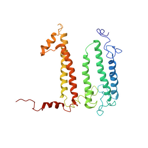
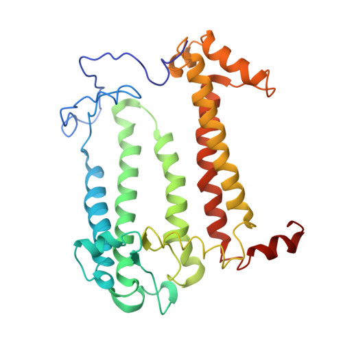
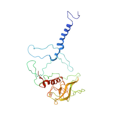
Structure of the membrane-bound protein photosynthetic reaction center from Rhodobacter sphaeroides.

Chang, C.H.el-Kabbani, O.Tiede, D.Norris, J.Schiffer, M.

(1991) Biochemistry 30: 5352-5360

  • DOI: https://doi.org/10.1021/bi00236a005
  • Primary Citation of Related Structures:  
    2RCR

  • PubMed Abstract: 

    The structure of the photosynthetic reaction center (RC) from Rhodobacter sphaeroides was determined at 3.1-A resolution by the molecular replacement method, using the Rhodopseudomonas viridis RC as the search structure. Atomic coordinates were refined with the difference Fourier method and restrained least-squares refinement techniques to a current R factor of 22%. The tertiary structure of the RC complex is stabilized by hydrophobic interactions between the L and M chains, by interactions of the pigments with each other and with the L and M chains, by residues from the L and M chains that coordinate to the Fe2+, by salt bridges that are formed between the L and M chains and the H chain, and possibly by electrostatic forces between the ends of helices. The conserved residues at the N-termini of the L and M chains were identified as recognition sites for the H chain.


  • Organizational Affiliation

    Biological and Medical Research Division, Argonne National Laboratory, Illinois 60439.


Macromolecules
Find similar proteins by:  (by identity cutoff)  |  3D Structure
Entity ID: 1
MoleculeChains Sequence LengthOrganismDetailsImage
PHOTOSYNTHETIC REACTION CENTER (L SUBUNIT)A [auth L]281Cereibacter sphaeroidesMutation(s): 0 
Membrane Entity: Yes 
UniProt
Find proteins for P0C0Y8 (Cereibacter sphaeroides)
Explore P0C0Y8 
Go to UniProtKB:  P0C0Y8
Entity Groups  
Sequence Clusters30% Identity50% Identity70% Identity90% Identity95% Identity100% Identity
UniProt GroupP0C0Y8
Sequence Annotations
Expand
  • Reference Sequence
Find similar proteins by:  (by identity cutoff)  |  3D Structure
Entity ID: 2
MoleculeChains Sequence LengthOrganismDetailsImage
PHOTOSYNTHETIC REACTION CENTER (M SUBUNIT)B [auth M]307Cereibacter sphaeroidesMutation(s): 0 
Membrane Entity: Yes 
UniProt
Find proteins for P0C0Y9 (Cereibacter sphaeroides)
Explore P0C0Y9 
Go to UniProtKB:  P0C0Y9
Entity Groups  
Sequence Clusters30% Identity50% Identity70% Identity90% Identity95% Identity100% Identity
UniProt GroupP0C0Y9
Sequence Annotations
Expand
  • Reference Sequence
Find similar proteins by:  (by identity cutoff)  |  3D Structure
Entity ID: 3
MoleculeChains Sequence LengthOrganismDetailsImage
PHOTOSYNTHETIC REACTION CENTER (H SUBUNIT)C [auth H]260Cereibacter sphaeroidesMutation(s): 0 
Membrane Entity: Yes 
UniProt
Find proteins for P0C0Y7 (Cereibacter sphaeroides)
Explore P0C0Y7 
Go to UniProtKB:  P0C0Y7
Entity Groups  
Sequence Clusters30% Identity50% Identity70% Identity90% Identity95% Identity100% Identity
UniProt GroupP0C0Y7
Sequence Annotations
Expand
  • Reference Sequence
Small Molecules
Ligands 4 Unique
IDChains Name / Formula / InChI Key2D Diagram3D Interactions
BCL
Query on BCL

Download Ideal Coordinates CCD File 
D [auth L],
E [auth L],
I [auth M],
J [auth M]
BACTERIOCHLOROPHYLL A
C55 H74 Mg N4 O6
DSJXIQQMORJERS-AGGZHOMASA-M
BPH
Query on BPH

Download Ideal Coordinates CCD File 
F [auth L],
K [auth M]
BACTERIOPHEOPHYTIN A
C55 H76 N4 O6
KWOZSBGNAHVCKG-SZQBJALDSA-N
UQ
Query on UQ

Download Ideal Coordinates CCD File 
G [auth L],
L [auth M]
Coenzyme Q10, (2Z,6E,10Z,14E,18E,22E,26Z)-isomer
C59 H90 O4
ACTIUHUUMQJHFO-RECDIHICSA-N
FE
Query on FE

Download Ideal Coordinates CCD File 
H [auth M]FE (III) ION
Fe
VTLYFUHAOXGGBS-UHFFFAOYSA-N
Experimental Data & Validation

Experimental Data

  • Method: X-RAY DIFFRACTION
  • Resolution: 3.10 Å
  • R-Value Observed: 0.220 
  • Space Group: P 21 21 21
Unit Cell:
Length ( Å )Angle ( ˚ )
a = 142.2α = 90
b = 139.6β = 90
c = 78.7γ = 90
Software Package:
Software NamePurpose
PROLSQrefinement

Structure Validation

View Full Validation Report


Entry History 

Deposition Data

Revision History  (Full details and data files)

  • Version 1.0: 1993-07-15
    Type: Initial release
  • Version 1.1: 2008-03-25
    Changes: Version format compliance
  • Version 1.2: 2011-07-13
    Changes: Non-polymer description, Version format compliance
  • Version 1.3: 2024-02-21
    Changes: Data collection, Database references, Derived calculations, Other