2QTV

Structure of Sec23-Sar1 complexed with the active fragment of Sec31


Experimental Data Snapshot

  • Method: X-RAY DIFFRACTION
  • Resolution: 2.50 Å
  • R-Value Free: 0.262 
  • R-Value Work: 0.205 
  • R-Value Observed: 0.208 

wwPDB Validation   3D Report Full Report


Ligand Structure Quality Assessment 


This is version 1.4 of the entry. See complete history


Literature

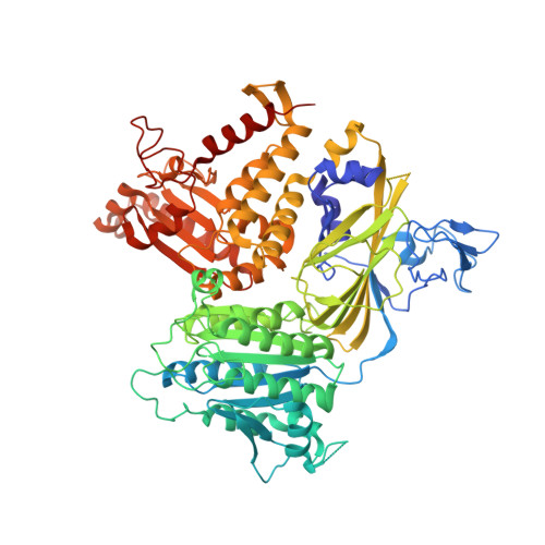
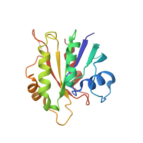
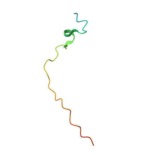
Insights into COPII coat nucleation from the structure of Sec23.Sar1 complexed with the active fragment of Sec31.

Bi, X.Mancias, J.D.Goldberg, J.

(2007) Dev Cell 13: 635-645

  • DOI: https://doi.org/10.1016/j.devcel.2007.10.006
  • Primary Citation of Related Structures:  
    2QTV

  • PubMed Abstract: 

    The COPII vesicular coat forms on the endoplasmic reticulum from Sar1-GTP, Sec23/24 and Sec13/31 protein subunits. Here, we define the interaction between Sec23/24.Sar1 and Sec13/31, involving a 40 residue Sec31 fragment. In the crystal structure of the ternary complex, Sec31 binds as an extended polypeptide across a composite surface of the Sec23 and Sar1-GTP molecules, explaining the stepwise character of Sec23/24.Sar1 and Sec13/31 recruitment to the membrane. The Sec31 fragment stimulates GAP activity of Sec23/24, and a convergence of Sec31 and Sec23 residues at the Sar1 GTPase active site explains how GTP hydrolysis is triggered leading to COPII coat disassembly. The Sec31 active fragment is accommodated in a binding groove supported in part by Sec23 residue Phe380. Substitution of the corresponding residue F382L in human Sec23A causes cranio-lenticulo-sutural dysplasia, and we suggest that this mutation disrupts the nucleation of COPII coat proteins at endoplasmic reticulum exit sites.


  • Organizational Affiliation

    Howard Hughes Medical Institute and the Structural Biology Program, Memorial Sloan-Kettering Cancer Center, 1275 York Avenue, New York, NY 10021, USA.


Macromolecules
Find similar proteins by:  (by identity cutoff)  |  3D Structure
Entity ID: 1
MoleculeChains Sequence LengthOrganismDetailsImage
Protein transport protein SEC23772Saccharomyces cerevisiaeMutation(s): 0 
Gene Names: SEC23
UniProt
Find proteins for P15303 (Saccharomyces cerevisiae (strain ATCC 204508 / S288c))
Explore P15303 
Go to UniProtKB:  P15303
Entity Groups  
Sequence Clusters30% Identity50% Identity70% Identity90% Identity95% Identity100% Identity
UniProt GroupP15303
Sequence Annotations
Expand
  • Reference Sequence
Find similar proteins by:  (by identity cutoff)  |  3D Structure
Entity ID: 2
MoleculeChains Sequence LengthOrganismDetailsImage
Small COPII coat GTPase SAR1167Saccharomyces cerevisiaeMutation(s): 0 
Gene Names: SAR1
EC: 3.6.5
UniProt
Find proteins for P20606 (Saccharomyces cerevisiae (strain ATCC 204508 / S288c))
Explore P20606 
Go to UniProtKB:  P20606
Entity Groups  
Sequence Clusters30% Identity50% Identity70% Identity90% Identity95% Identity100% Identity
UniProt GroupP20606
Sequence Annotations
Expand
  • Reference Sequence
Find similar proteins by:  (by identity cutoff)  |  3D Structure
Entity ID: 3
MoleculeChains Sequence LengthOrganismDetailsImage
Protein transport protein SEC31C [auth D]49Saccharomyces cerevisiaeMutation(s): 0 
Gene Names: SEC31WEB1
UniProt
Find proteins for P38968 (Saccharomyces cerevisiae (strain ATCC 204508 / S288c))
Explore P38968 
Go to UniProtKB:  P38968
Entity Groups  
Sequence Clusters30% Identity50% Identity70% Identity90% Identity95% Identity100% Identity
UniProt GroupP38968
Sequence Annotations
Expand
  • Reference Sequence
Experimental Data & Validation

Experimental Data

  • Method: X-RAY DIFFRACTION
  • Resolution: 2.50 Å
  • R-Value Free: 0.262 
  • R-Value Work: 0.205 
  • R-Value Observed: 0.208 
  • Space Group: P 21 21 2
Unit Cell:
Length ( Å )Angle ( ˚ )
a = 94.748α = 90
b = 133.213β = 90
c = 82.041γ = 90
Software Package:
Software NamePurpose
DENZOdata reduction
SCALEPACKdata scaling
CNSrefinement
PDB_EXTRACTdata extraction
HKL-2000data collection
AMoREphasing

Structure Validation

View Full Validation Report


Ligand Structure Quality Assessment 


Entry History 

Deposition Data

Revision History  (Full details and data files)

  • Version 1.0: 2008-01-15
    Type: Initial release
  • Version 1.1: 2011-07-13
    Changes: Version format compliance
  • Version 1.2: 2017-10-25
    Changes: Refinement description
  • Version 1.3: 2018-02-14
    Changes: Experimental preparation
  • Version 1.4: 2024-02-21
    Changes: Data collection, Database references, Derived calculations