2Q6W

The structure of HLA-DRA, DRB3*0101 (DR52a) with bound platelet integrin peptide associated with fetal and neonatal alloimmune thrombocytopenia


Experimental Data Snapshot

  • Method: X-RAY DIFFRACTION
  • Resolution: 2.25 Å
  • R-Value Free: 0.265 
  • R-Value Work: 0.210 
  • R-Value Observed: 0.214 

wwPDB Validation   3D Report Full Report


This is version 1.4 of the entry. See complete history


Literature

Crystallographic Structure of the Human Leukocyte Antigen DRA, DRB3*0101: Models of a Directional Alloimmune Response and Autoimmunity

Parry, C.S.Gorski, J.Stern, L.J.

(2007) J Mol Biol 371: 435-446

  • DOI: https://doi.org/10.1016/j.jmb.2007.05.025
  • Primary Citation of Related Structures:  
    2Q6W

  • PubMed Abstract: 

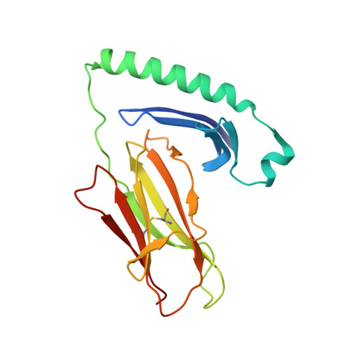
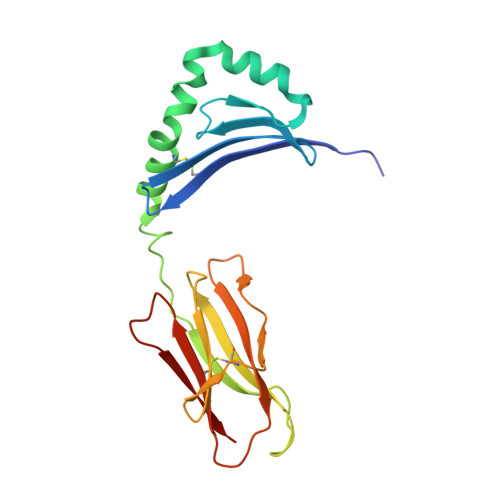
    We describe structural studies of the human leukocyte antigen DR52a, HLA-DRA/DRB3*0101, in complex with an N-terminal human platelet integrin alphaII(B)betaIII glycoprotein peptide which contains a Leu/Pro dimorphism. The 33:Leu dimorphism is the epitope for the T cell directed response in neonatal alloimmune thrombocytopenia and post-transfusion purpura in individuals with the alphaII(B)betaIII 33:Pro allele, and defines the unidirectional alloimmune response. This condition is always associated with DR52a. The crystallographic structure has been refined to 2.25 A. There are two alphabeta heterodimers to the asymmetric unit in space group P4(1)2(1)2. The molecule is characterized by two prominent hydrophobic pockets at either end of the peptide binding cleft and a deep, narrower and highly charged P4 opening underneath the beta 1 chain. Further, the peptide in the second molecule displays a sharp upward turn after pocket P9. The structure reveals the role of pockets and the distinctive basic P4 pocket, shared by DR52a and DR3, in selecting their respective binding peptide repertoire. We observe an interesting switch in a residue from the canonically assigned pocket 6 seen in prior class II structures to pocket 4. This occludes the P6 pocket helping to explain the distinctive "1-4-9" peptide binding motif. A beta57 Asp-->Val substitution abrogates the salt-bridge to alpha76 Arg and along with a hydrophobic beta37 is important in shaping the P9 pocket. DRB3*0101 and DRB1*0301 belong to an ancestral haplotype and are associated with many autoimmune diseases linked to antigen presentation, but whereas DR3 is susceptible to type 1 diabetes DR52a is not. This dichotomy is explored for clues to the disease.


  • Organizational Affiliation

    Massachusetts Institute of Technology, Department of Chemistry, 77 Massachusetts Avenue, Cambridge, MA 02139, USA. csparry@helix.nih.gov


Macromolecules
Find similar proteins by:  (by identity cutoff)  |  3D Structure
Entity ID: 1
MoleculeChains Sequence LengthOrganismDetailsImage
HLA class II histocompatibility antigen, DR alpha chain
A, D
182Homo sapiensMutation(s): 0 
Gene Names: HLA-DRA
UniProt & NIH Common Fund Data Resources
Find proteins for P01903 (Homo sapiens)
Explore P01903 
Go to UniProtKB:  P01903
PHAROS:  P01903
GTEx:  ENSG00000204287 
Entity Groups  
Sequence Clusters30% Identity50% Identity70% Identity90% Identity95% Identity100% Identity
UniProt GroupP01903
Sequence Annotations
Expand
  • Reference Sequence
Find similar proteins by:  (by identity cutoff)  |  3D Structure
Entity ID: 2
MoleculeChains Sequence LengthOrganismDetailsImage
HLA class II histocompatibility antigen, DRB3-1 beta chain
B, E
190Homo sapiensMutation(s): 0 
Gene Names: HLA-DRB3
UniProt
Find proteins for P79483 (Homo sapiens)
Explore P79483 
Go to UniProtKB:  P79483
Entity Groups  
Sequence Clusters30% Identity50% Identity70% Identity90% Identity95% Identity100% Identity
UniProt GroupP79483
Sequence Annotations
Expand
  • Reference Sequence

Find similar proteins by:  Sequence   |   3D Structure  

Entity ID: 3
MoleculeChains Sequence LengthOrganismDetailsImage
Integrin beta-3
C, F
12Homo sapiensMutation(s): 1 
UniProt & NIH Common Fund Data Resources
Find proteins for P05106 (Homo sapiens)
Explore P05106 
Go to UniProtKB:  P05106
PHAROS:  P05106
GTEx:  ENSG00000259207 
Entity Groups  
Sequence Clusters30% Identity50% Identity70% Identity90% Identity95% Identity100% Identity
UniProt GroupP05106
Sequence Annotations
Expand
  • Reference Sequence
Experimental Data & Validation

Experimental Data

  • Method: X-RAY DIFFRACTION
  • Resolution: 2.25 Å
  • R-Value Free: 0.265 
  • R-Value Work: 0.210 
  • R-Value Observed: 0.214 
  • Space Group: P 41 21 2
Unit Cell:
Length ( Å )Angle ( ˚ )
a = 92.169α = 90
b = 92.169β = 90
c = 248.555γ = 90
Software Package:
Software NamePurpose
REFMACrefinement
HKL-2000data collection
HKL-2000data reduction
HKL-2000data scaling
AMoREphasing

Structure Validation

View Full Validation Report


Entry History 

Deposition Data

  • Released Date: 2007-07-24 
  • Deposition Author(s): Parry, C.S.

Revision History  (Full details and data files)

  • Version 1.0: 2007-07-24
    Type: Initial release
  • Version 1.1: 2008-05-01
    Changes: Version format compliance
  • Version 1.2: 2011-07-13
    Changes: Version format compliance
  • Version 1.3: 2021-10-20
    Changes: Database references, Source and taxonomy
  • Version 1.4: 2023-08-30
    Changes: Data collection, Refinement description