2IUF

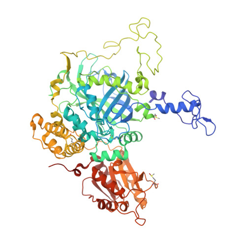
The structures of Penicillium vitale catalase: resting state, oxidised state (compound I) and complex with aminotriazole


Experimental Data Snapshot

  • Method: X-RAY DIFFRACTION
  • Resolution: 1.71 Å
  • R-Value Free: 0.154 
  • R-Value Work: 0.129 
  • R-Value Observed: 0.131 

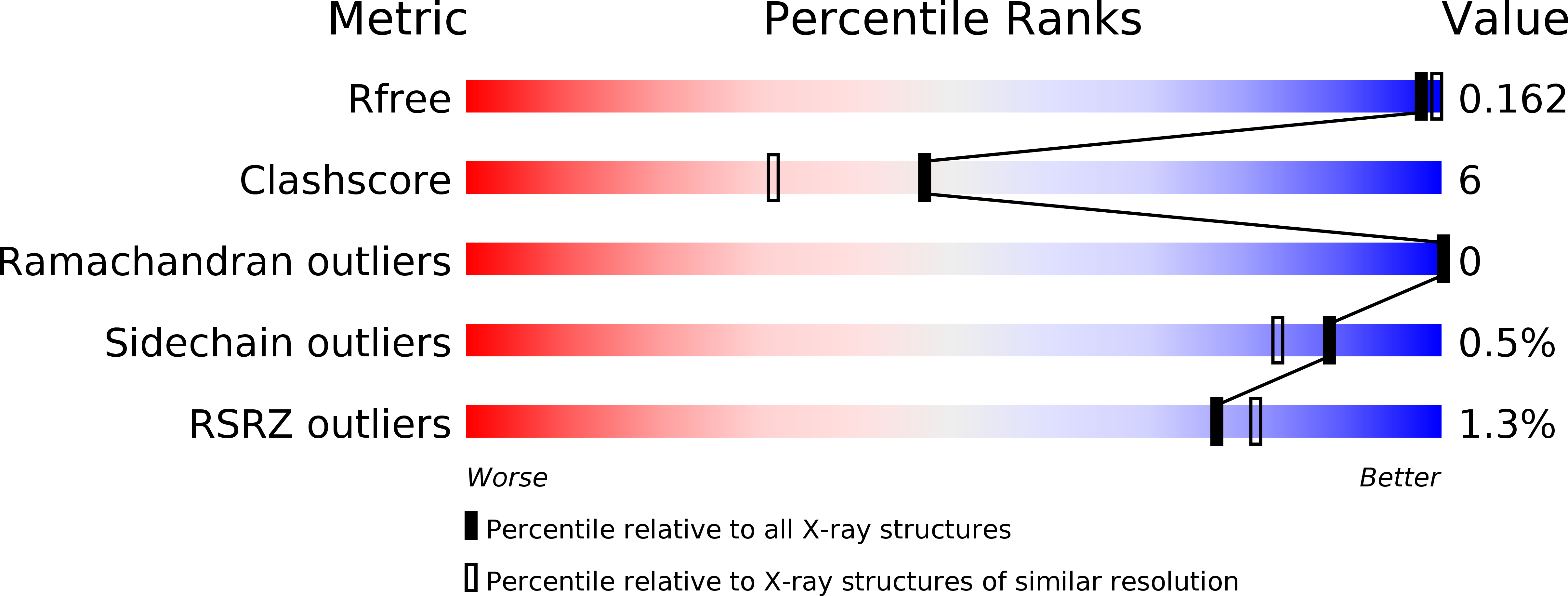
wwPDB Validation   3D Report Full Report


Ligand Structure Quality Assessment 


This is version 2.0 of the entry. See complete history


Literature

The Structures and Electronic Configuration of Compound I Intermediates of Helicobacter Pylori and Penicillium Vitale Catalases Determined by X-Ray Crystallography and Qm/Mm Density Functional Theory Calculations.

Alfonso-Prieto, M.Borovik, A.Carpena, X.Murshudov, G.Melik-Adamyan, W.Fita, I.Rovira, C.Loewen, P.C.

(2007) J Am Chem Soc 129: 4193

  • DOI: https://doi.org/10.1021/ja063660y
  • Primary Citation of Related Structures:  
    2IQF, 2IUF

  • PubMed Abstract: 

    The structures of Helicobacter pylori (HPC) and Penicillium vitale (PVC) catalases, each with two subunits in the crystal asymmetric unit, oxidized with peroxoacetic acid are reported at 1.8 and 1.7 A resolution, respectively. Despite the similar oxidation conditions employed, the iron-oxygen coordination length is 1.72 A for PVC, close to what is expected for a Fe=O double bond, and 1.80 and 1.85 A for HPC, suggestive of a Fe-O single bond. The structure and electronic configuration of the oxoferryl heme and immediate protein environment is investigated further by QM/MM density functional theory calculations. Four different active site electronic configurations are considered, Por*+-FeIV=O, Por*+-FeIV=O...HisH+, Por*+-FeIV-OH+ and Por-FeIV-OH (a protein radical is assumed in the latter configuration). The electronic structure of the primary oxidized species, Por*+-FeIV=O, differs qualitatively between HPC and PVC with an A2u-like porphyrin radical delocalized on the porphyrin in HPC and a mixed A1u-like "fluctuating" radical partially delocalized over the essential distal histidine, the porphyrin, and, to a lesser extent, the proximal tyrosine residue. This difference is rationalized in terms of HPC containing heme b and PVC containing heme d. It is concluded that compound I of PVC contains an oxoferryl Por*+-FeIV=O species with partial protonation of the distal histidine and compound I of HPC contains a hydroxoferryl Por-FeIV-OH with the second oxidation equivalent delocalized as a protein radical. The findings support the idea that there is a relation between radical migration to the protein and protonation of the oxoferryl bond in catalase.


  • Organizational Affiliation

    Centre especial de Recerca en Química Teorica, Parc Científic de Barcelona, Josep Samitier 1-5, 08028 Barcelona, Spain.


Macromolecules
Find similar proteins by:  (by identity cutoff)  |  3D Structure
Entity ID: 1
MoleculeChains Sequence LengthOrganismDetailsImage
CATALASEA,
B [auth E]
688Penicillium janthinellumMutation(s): 0 
EC: 1.11.1.6
UniProt
Find proteins for D9N167 (Penicillium janthinellum)
Explore D9N167 
Go to UniProtKB:  D9N167
Entity Groups  
Sequence Clusters30% Identity50% Identity70% Identity90% Identity95% Identity100% Identity
UniProt GroupD9N167
Sequence Annotations
Expand
  • Reference Sequence
Oligosaccharides

Help

Entity ID: 2
MoleculeChains Length2D Diagram Glycosylation3D Interactions
2-acetamido-2-deoxy-beta-D-glucopyranose-(1-6)-2-acetamido-2-deoxy-beta-D-glucopyranoseC [auth B]2N-Glycosylation
Glycosylation Resources
GlyTouCan:  G35932AR
GlyCosmos:  G35932AR
GlyGen:  G35932AR
Small Molecules
Ligands 7 Unique
IDChains Name / Formula / InChI Key2D Diagram3D Interactions
HDD
Query on HDD

Download Ideal Coordinates CCD File 
D [auth A],
R [auth E]
CIS-HEME D HYDROXYCHLORIN GAMMA-SPIROLACTONE
C34 H32 Fe N4 O5
UMGOPAWIGKFTRK-QQDQPIDJSA-N
NAG
Query on NAG

Download Ideal Coordinates CCD File 
E [auth A],
F [auth A],
G [auth A],
S [auth E],
T [auth E]
2-acetamido-2-deoxy-beta-D-glucopyranose
C8 H15 N O6
OVRNDRQMDRJTHS-FMDGEEDCSA-N
MPD
Query on MPD

Download Ideal Coordinates CCD File 
Q [auth A](4S)-2-METHYL-2,4-PENTANEDIOL
C6 H14 O2
SVTBMSDMJJWYQN-YFKPBYRVSA-N
F50
Query on F50

Download Ideal Coordinates CCD File 
DA [auth E],
L [auth A],
Y [auth E]
ETHANEPEROXOIC ACID
C2 H4 O3
KFSLWBXXFJQRDL-UHFFFAOYSA-N
ACT
Query on ACT

Download Ideal Coordinates CCD File 
AA [auth E]
BA [auth E]
CA [auth E]
EA [auth E]
FA [auth E]
AA [auth E],
BA [auth E],
CA [auth E],
EA [auth E],
FA [auth E],
IA [auth E],
K [auth A],
M [auth A],
N [auth A],
O [auth A],
P [auth A],
X [auth E],
Z [auth E]
ACETATE ION
C2 H3 O2
QTBSBXVTEAMEQO-UHFFFAOYSA-M
CA
Query on CA

Download Ideal Coordinates CCD File 
GA [auth E]
HA [auth E]
I [auth A]
J [auth A]
V [auth E]
GA [auth E],
HA [auth E],
I [auth A],
J [auth A],
V [auth E],
W [auth E]
CALCIUM ION
Ca
BHPQYMZQTOCNFJ-UHFFFAOYSA-N
O
Query on O

Download Ideal Coordinates CCD File 
H [auth A],
U [auth E]
OXYGEN ATOM
O
XLYOFNOQVPJJNP-UHFFFAOYSA-N
Modified Residues  1 Unique
IDChains TypeFormula2D DiagramParent
MHO
Query on MHO
A,
B [auth E]
L-PEPTIDE LINKINGC5 H11 N O3 SMET
Experimental Data & Validation

Experimental Data

  • Method: X-RAY DIFFRACTION
  • Resolution: 1.71 Å
  • R-Value Free: 0.154 
  • R-Value Work: 0.129 
  • R-Value Observed: 0.131 
  • Space Group: P 31 2 1
Unit Cell:
Length ( Å )Angle ( ˚ )
a = 142.44α = 90
b = 142.44β = 90
c = 132.23γ = 120
Software Package:
Software NamePurpose
REFMACrefinement
DENZOdata reduction
SCALEPACKdata scaling

Structure Validation

View Full Validation Report


Ligand Structure Quality Assessment 


Entry History 

Deposition Data

Revision History  (Full details and data files)

  • Version 1.0: 2006-07-10
    Type: Initial release
  • Version 1.1: 2016-12-28
    Changes: Database references, Derived calculations, Non-polymer description, Other, Structure summary, Version format compliance
  • Version 2.0: 2020-07-29
    Type: Remediation
    Reason: Carbohydrate remediation
    Changes: Advisory, Atomic model, Data collection, Derived calculations, Other, Structure summary