2IH9

A high-dose crystal structure of a recombinant Melanocarbus albomyces laccase


Experimental Data Snapshot

  • Method: X-RAY DIFFRACTION
  • Resolution: 2.00 Å
  • R-Value Free: 0.299 
  • R-Value Work: 0.263 
  • R-Value Observed: 0.263 

wwPDB Validation   3D Report Full Report


Ligand Structure Quality Assessment 


This is version 2.1 of the entry. See complete history


Literature

A crystallographic and spectroscopic study on the effect of X-ray radiation on the crystal structure of Melanocarpus albomyces laccase.

Hakulinen, N.Kruus, K.Koivula, A.Rouvinen, J.

(2006) Biochem Biophys Res Commun 350: 929-934

  • DOI: https://doi.org/10.1016/j.bbrc.2006.09.144
  • Primary Citation of Related Structures:  
    2IH8, 2IH9

  • PubMed Abstract: 

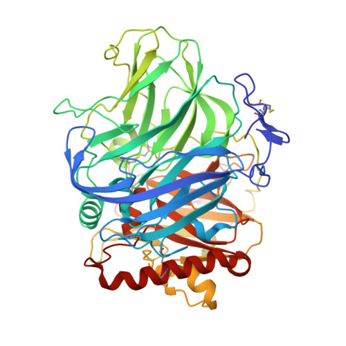
    Laccases (p-diphenol dioxygen oxidoreductases) belong to the family of blue multicopper oxidases, which catalyse the four-electron reduction of dioxygen to water concomitantly through the oxidation of substrate molecules. Blue multicopper oxidases have four coppers, a copper (T1) forming a mononuclear site and a cluster of three coppers (T2, T3, and T3') forming a trinuclear site. Because X-rays are known to liberate electrons during data collection and may thus affect the oxidation state of metals, we have investigated the effect of X-ray radiation upon the crystal structure of a recombinant laccase from Melanocarpus albomyces through the use of crystallography and crystal absorption spectroscopy. Two data sets with different strategies, a low and a high-dose data set, were collected at synchrotron. We have observed earlier that the trinuclear site had an elongated electron density amidst coppers, suggesting dioxygen binding. The low-dose synchrotron structure showed similar elongated electron density, but the high-dose X-ray radiation removed the bulk of this density. Therefore, X-ray radiation could alter the active site of laccase from M. albomyces. Absorption spectra of the crystals (320, 420, and 590nm) during X-ray radiation were measured at a home laboratory. Spectra clearly showed how that the band at 590nm had vanished, resulting from the T1 copper being reduced, during the long X-ray measurements. The crystal colour changed from blue to colourless. Absorptions at 320 and 420nm seemed to be rather permanent. The absorption at 320nm is due to the T3 coppers and it is proposed that absorption at 420nm is due to the T2 copper when dioxygen or a reaction intermediate is close to this copper.


  • Organizational Affiliation

    Department of Chemistry, University of Joensuu, P.O. Box 111, FIN-80101 Joensuu, Finland. nina.hakulinen@joensuu.fi


Macromolecules
Find similar proteins by:  (by identity cutoff)  |  3D Structure
Entity ID: 1
MoleculeChains Sequence LengthOrganismDetailsImage
Laccase-1
A, B
559Melanocarpus albomycesMutation(s): 0 
Gene Names: LAC1
EC: 1.10.3.2
UniProt
Find proteins for Q70KY3 (Melanocarpus albomyces)
Explore Q70KY3 
Go to UniProtKB:  Q70KY3
Entity Groups  
Sequence Clusters30% Identity50% Identity70% Identity90% Identity95% Identity100% Identity
UniProt GroupQ70KY3
Sequence Annotations
Expand
  • Reference Sequence
Oligosaccharides

Help

Entity ID: 2
MoleculeChains Length2D Diagram Glycosylation3D Interactions
beta-D-mannopyranose-(1-4)-2-acetamido-2-deoxy-beta-D-glucopyranose-(1-4)-2-acetamido-2-deoxy-beta-D-glucopyranose
C, D, F, G
3N-Glycosylation
Glycosylation Resources
GlyTouCan:  G15407YE
GlyCosmos:  G15407YE
GlyGen:  G15407YE
Entity ID: 3
MoleculeChains Length2D Diagram Glycosylation3D Interactions
2-acetamido-2-deoxy-beta-D-glucopyranose-(1-4)-2-acetamido-2-deoxy-beta-D-glucopyranose
E, H
2N-Glycosylation
Glycosylation Resources
GlyTouCan:  G42666HT
GlyCosmos:  G42666HT
GlyGen:  G42666HT
Small Molecules
Ligands 4 Unique
IDChains Name / Formula / InChI Key2D Diagram3D Interactions
NAG
Query on NAG

Download Ideal Coordinates CCD File 
EA [auth B]
I [auth A]
J [auth A]
K [auth A]
L [auth A]
EA [auth B],
I [auth A],
J [auth A],
K [auth A],
L [auth A],
T [auth A],
U [auth B],
V [auth B],
W [auth B],
X [auth B]
2-acetamido-2-deoxy-beta-D-glucopyranose
C8 H15 N O6
OVRNDRQMDRJTHS-FMDGEEDCSA-N
SO4
Query on SO4

Download Ideal Coordinates CCD File 
DA [auth B],
R [auth A],
S [auth A]
SULFATE ION
O4 S
QAOWNCQODCNURD-UHFFFAOYSA-L
CU
Query on CU

Download Ideal Coordinates CCD File 
AA [auth B]
BA [auth B]
M [auth A]
N [auth A]
O [auth A]
AA [auth B],
BA [auth B],
M [auth A],
N [auth A],
O [auth A],
P [auth A],
Y [auth B],
Z [auth B]
COPPER (II) ION
Cu
JPVYNHNXODAKFH-UHFFFAOYSA-N
CL
Query on CL

Download Ideal Coordinates CCD File 
CA [auth B],
Q [auth A]
CHLORIDE ION
Cl
VEXZGXHMUGYJMC-UHFFFAOYSA-M
Experimental Data & Validation

Experimental Data

  • Method: X-RAY DIFFRACTION
  • Resolution: 2.00 Å
  • R-Value Free: 0.299 
  • R-Value Work: 0.263 
  • R-Value Observed: 0.263 
  • Space Group: C 1 2 1
Unit Cell:
Length ( Å )Angle ( ˚ )
a = 174.49α = 90
b = 62.33β = 96.25
c = 125.1γ = 90
Software Package:
Software NamePurpose
XSCALEdata processing
CNSrefinement
PDB_EXTRACTdata extraction
XDSdata reduction
XSCALEdata scaling

Structure Validation

View Full Validation Report


Ligand Structure Quality Assessment 


Entry History 

Deposition Data

Revision History  (Full details and data files)

  • Version 1.0: 2006-11-14
    Type: Initial release
  • Version 1.1: 2008-05-01
    Changes: Version format compliance
  • Version 1.2: 2011-07-13
    Changes: Non-polymer description, Version format compliance
  • Version 1.3: 2016-08-17
    Changes: Non-polymer description
  • Version 2.0: 2020-07-29
    Type: Remediation
    Reason: Carbohydrate remediation
    Changes: Advisory, Atomic model, Data collection, Derived calculations, Structure summary
  • Version 2.1: 2023-08-30
    Changes: Data collection, Database references, Refinement description, Structure summary