2HOR

Crystal structure of alliinase from garlic- apo form


Experimental Data Snapshot

  • Method: X-RAY DIFFRACTION
  • Resolution: 1.60 Å
  • R-Value Free: 0.179 
  • R-Value Work: 0.163 
  • R-Value Observed: 0.164 

wwPDB Validation   3D Report Full Report


This is version 2.0 of the entry. See complete history


Literature

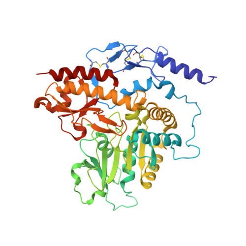
Two Structures of Alliinase from Alliium sativum L.: Apo Form and Ternary Complex with Aminoacrylate Reaction Intermediate Covalently Bound to the PLP Cofactor.

Shimon, L.J.Rabinkov, A.Shin, I.Miron, T.Mirelman, D.Wilchek, M.Frolow, F.

(2007) J Mol Biol 366: 611-625

  • DOI: https://doi.org/10.1016/j.jmb.2006.11.041
  • Primary Citation of Related Structures:  
    2HOR, 2HOX

  • PubMed Abstract: 

    Alliinase (alliin lyase EC 4.4.1.4), a PLP-dependent alpha, beta-eliminating lyase, constitutes one of the major protein components of garlic (Alliium sativum L.) bulbs. The enzyme is a homodimeric glycoprotein and catalyzes the conversion of a specific non-protein sulfur-containing amino acid alliin ((+S)-allyl-L-cysteine sulfoxide) to allicin (diallyl thiosulfinate, the well known biologically active component of freshly crushed garlic), pyruvate and ammonia. The enzyme was crystallized in the presence of (+S)-allyl-L-cysteine, forming dendrite-like monoclinic crystals. In addition, intentionally produced apo-enzyme was crystallized in tetragonal form. These structures of alliinase with associated glycans were resolved to 1.4 A and 1.61 A by molecular replacement. Branched hexasaccharide chains N-linked to Asn146 and trisaccharide chains N-linked to Asn328 are seen. The structure of hexasaccharide was found similar to "short chain complex vacuole type" oligosaccharide most commonly seen in plant glycoproteins. An unexpected state of the enzyme active site has been observed in the present structure. The electron density in the region of the cofactor made it possible to identify the cofactor moiety as aminoacrylate intermediate covalently bound to the PLP cofactor. It was found in the present structure to be stabilized by large number of interactions with surrounding protein residues. Moreover, the existence of the expected internal aldimine bond between the epsilon-amino group of Lys251 and the aldehyde of the PLP is ruled out on the basis of a distinct separation of electron density of Lys251. The structure of the active site cavity in the apo-form is nearly identical to that seen in the holo-form, with two sulfate ions, an acetate and several water molecules from crystallization conditions that replace and mimic the PLP cofactor.


  • Organizational Affiliation

    Department of Chemical Research Support, The Weizmann Institute of Science, Rehovot 76100, Israel.


Macromolecules
Find similar proteins by:  (by identity cutoff)  |  3D Structure
Entity ID: 1
MoleculeChains Sequence LengthOrganismDetailsImage
Alliin lyase 1427Allium sativumMutation(s): 0 
EC: 4.4.1.4
UniProt
Find proteins for Q01594 (Allium sativum)
Explore Q01594 
Go to UniProtKB:  Q01594
Entity Groups  
Sequence Clusters30% Identity50% Identity70% Identity90% Identity95% Identity100% Identity
UniProt GroupQ01594
Sequence Annotations
Expand
  • Reference Sequence
Oligosaccharides

Help

Entity ID: 2
MoleculeChains Length2D Diagram Glycosylation3D Interactions
alpha-D-mannopyranose-(2-3)-[alpha-D-mannopyranose-(1-6)]beta-D-mannopyranose-(1-4)-2-acetamido-2-deoxy-beta-D-glucopyranose-(1-4)-[alpha-L-fucopyranose-(1-3)]2-acetamido-2-deoxy-beta-D-glucopyranose
B
6N/AN-Glycosylation
Small Molecules
Ligands 5 Unique
IDChains Name / Formula / InChI Key2D Diagram3D Interactions
NAG
Query on NAG

Download Ideal Coordinates CCD File 
C [auth A]2-acetamido-2-deoxy-beta-D-glucopyranose
C8 H15 N O6
OVRNDRQMDRJTHS-FMDGEEDCSA-N
SO4
Query on SO4

Download Ideal Coordinates CCD File 
D [auth A],
E [auth A],
G [auth A],
H [auth A],
I [auth A]
SULFATE ION
O4 S
QAOWNCQODCNURD-UHFFFAOYSA-L
NO3
Query on NO3

Download Ideal Coordinates CCD File 
L [auth A]NITRATE ION
N O3
NHNBFGGVMKEFGY-UHFFFAOYSA-N
ACT
Query on ACT

Download Ideal Coordinates CCD File 
F [auth A]ACETATE ION
C2 H3 O2
QTBSBXVTEAMEQO-UHFFFAOYSA-M
CL
Query on CL

Download Ideal Coordinates CCD File 
J [auth A],
K [auth A]
CHLORIDE ION
Cl
VEXZGXHMUGYJMC-UHFFFAOYSA-M
Experimental Data & Validation

Experimental Data

  • Method: X-RAY DIFFRACTION
  • Resolution: 1.60 Å
  • R-Value Free: 0.179 
  • R-Value Work: 0.163 
  • R-Value Observed: 0.164 
  • Space Group: P 41 21 2
Unit Cell:
Length ( Å )Angle ( ˚ )
a = 81.118α = 90
b = 81.118β = 90
c = 164.017γ = 90
Software Package:
Software NamePurpose
DENZOdata reduction
SCALEPACKdata scaling
REFMACrefinement
PDB_EXTRACTdata extraction
HKL-2000data reduction
MOLREPphasing

Structure Validation

View Full Validation Report


Entry History 

Deposition Data

Revision History  (Full details and data files)

  • Version 1.0: 2007-02-06
    Type: Initial release
  • Version 1.1: 2008-05-01
    Changes: Version format compliance
  • Version 1.2: 2011-07-13
    Changes: Derived calculations, Version format compliance
  • Version 1.3: 2014-04-16
    Changes: Other
  • Version 1.4: 2017-10-18
    Changes: Advisory, Refinement description
  • Version 1.5: 2018-01-24
    Changes: Structure summary
  • Version 2.0: 2020-07-29
    Type: Remediation
    Reason: Carbohydrate remediation
    Changes: Advisory, Atomic model, Data collection, Derived calculations, Structure summary