2GDU

E232Q mutant of sucrose phosphorylase from BIFIDOBACTERIUM ADOLESCENTIS in complex with sucrose


Experimental Data Snapshot

  • Method: X-RAY DIFFRACTION
  • Resolution: 2.10 Å
  • R-Value Free: 0.217 
  • R-Value Work: 0.182 
  • R-Value Observed: 0.184 

wwPDB Validation   3D Report Full Report


This is version 2.0 of the entry. See complete history


Literature

Structural Rearrangements of Sucrose Phosphorylase from Bifidobacterium adolescentis during Sucrose Conversion

Mirza, O.Skov, L.K.Sprogoe, D.van den Broek, L.A.M.Beldman, G.Kastrup, J.S.Gajhede, M.

(2006) J Biol Chem 281: 35576-35584

  • DOI: https://doi.org/10.1074/jbc.M605611200
  • Primary Citation of Related Structures:  
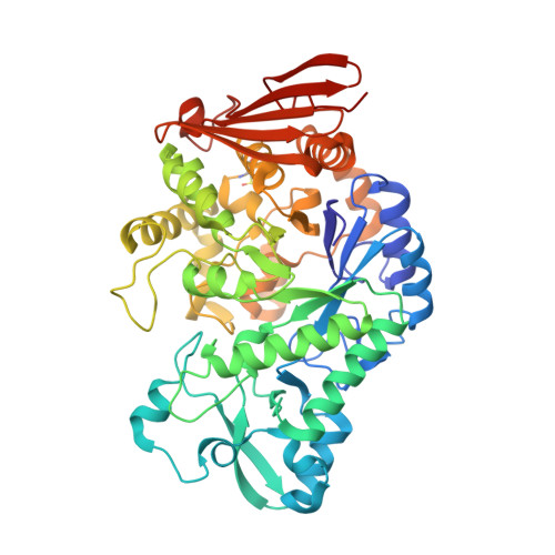
    2GDU

  • PubMed Abstract: 

    The reaction mechanism of sucrose phosphorylase from Bifidobacterium adolescentis (BiSP) was studied by site-directed mutagenesis and x-ray crystallography. An inactive mutant of BiSP (E232Q) was co-crystallized with sucrose. The structure revealed a substrate-binding mode comparable with that seen in other related sucrose-acting enzymes. Wild-type BiSP was also crystallized in the presence of sucrose. In the dimeric structure, a covalent glucosyl intermediate was formed in one molecule of the BiSP dimer, and after hydrolysis of the glucosyl intermediate, a beta-D-glucose product complex was formed in the other molecule. Although the overall structure of the BiSP-glucosyl intermediate complex is similar to that of the BiSP(E232Q)-sucrose complex, the glucose complex discloses major differences in loop conformations. Two loops (residues 336-344 and 132-137) in the proximity of the active site move up to 16 and 4 A, respectively. On the basis of these findings, we have suggested a reaction cycle that takes into account the large movements in the active-site entrance loops.


  • Organizational Affiliation

    Biostructural Research Unit, Department of Medicinal Chemistry, Danish University of Pharmaceutical Sciences, Universitetsparken 2, DK-2100 Copenhagen, Denmark.


Macromolecules
Find similar proteins by:  (by identity cutoff)  |  3D Structure
Entity ID: 1
MoleculeChains Sequence LengthOrganismDetailsImage
sucrose phosphorylase
A, B
504Bifidobacterium adolescentisMutation(s): 1 
EC: 2.4.1.7
UniProt
Find proteins for A0ZZH6 (Bifidobacterium adolescentis (strain ATCC 15703 / DSM 20083 / NCTC 11814 / E194a))
Explore A0ZZH6 
Go to UniProtKB:  A0ZZH6
Entity Groups  
Sequence Clusters30% Identity50% Identity70% Identity90% Identity95% Identity100% Identity
UniProt GroupA0ZZH6
Sequence Annotations
Expand
  • Reference Sequence
Oligosaccharides

Help

Entity ID: 2
MoleculeChains Length2D Diagram Glycosylation3D Interactions
beta-D-fructofuranose-(2-1)-alpha-D-glucopyranose
C, D
2N/A
Glycosylation Resources
GlyTouCan:  G05551OP
GlyCosmos:  G05551OP
Small Molecules
Modified Residues  1 Unique
IDChains TypeFormula2D DiagramParent
CSD
Query on CSD
A, B
L-PEPTIDE LINKINGC3 H7 N O4 SCYS
Biologically Interesting Molecules (External Reference) 1 Unique
Experimental Data & Validation

Experimental Data

  • Method: X-RAY DIFFRACTION
  • Resolution: 2.10 Å
  • R-Value Free: 0.217 
  • R-Value Work: 0.182 
  • R-Value Observed: 0.184 
  • Space Group: P 21 21 21
Unit Cell:
Length ( Å )Angle ( ˚ )
a = 55.331α = 90
b = 124.157β = 90
c = 152.453γ = 90
Software Package:
Software NamePurpose
CNSrefinement
PDB_EXTRACTdata extraction
SCALEPACKdata scaling
CNSphasing

Structure Validation

View Full Validation Report


Entry History 

Deposition Data

Revision History  (Full details and data files)

  • Version 1.0: 2006-09-26
    Type: Initial release
  • Version 1.1: 2008-05-01
    Changes: Version format compliance
  • Version 1.2: 2011-07-13
    Changes: Version format compliance
  • Version 1.3: 2014-11-12
    Changes: Other
  • Version 2.0: 2020-07-29
    Type: Remediation
    Reason: Carbohydrate remediation
    Changes: Atomic model, Data collection, Database references, Derived calculations, Non-polymer description, Structure summary