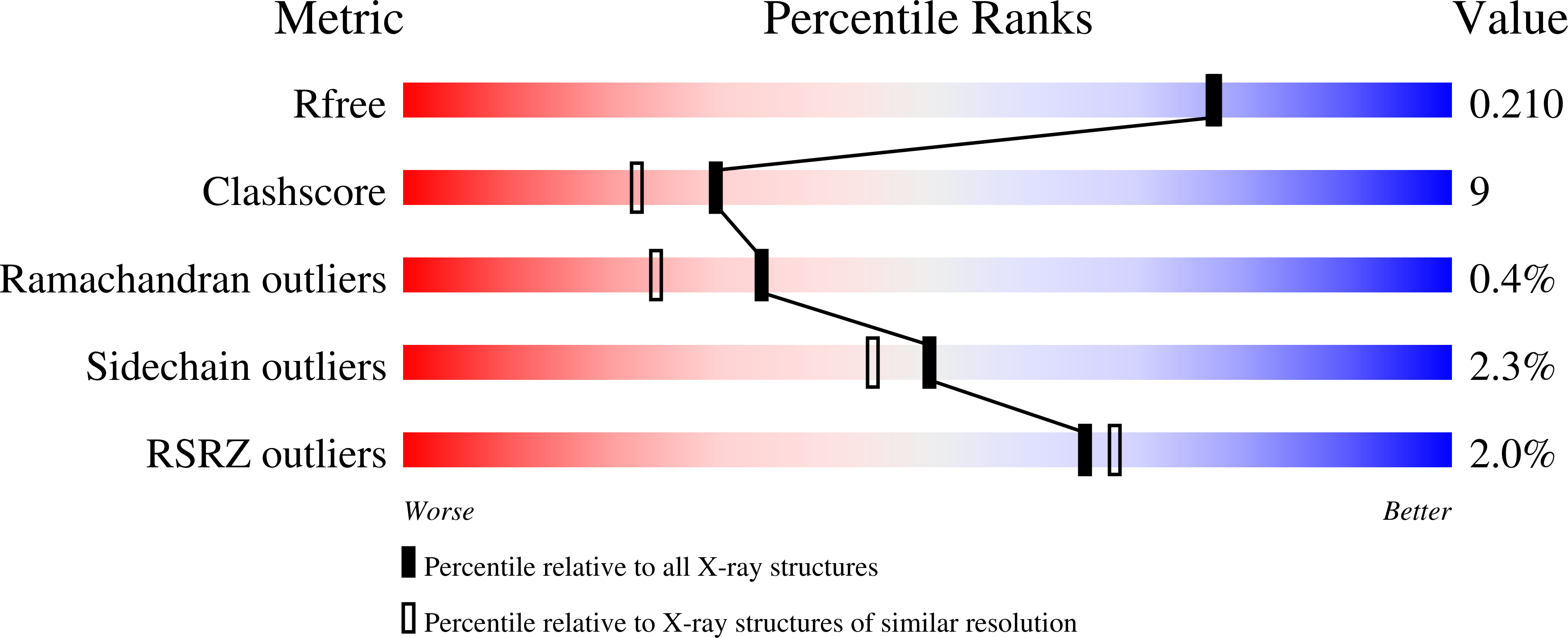
2FYV

Golgi alpha-mannosidase II complex with an amino-salacinol carboxylate analog


Experimental Data Snapshot

  • Method: X-RAY DIFFRACTION
  • Resolution: 1.90 Å
  • R-Value Free: 0.212 
  • R-Value Work: 0.162 
  • R-Value Observed: 0.162 

wwPDB Validation   3D Report Full Report


Ligand Structure Quality Assessment 


This is version 1.5 of the entry. See complete history


Literature

Synthesis, enzymatic activity, and X-ray crystallography of an unusual class of amino acids.

Chen, W.Kuntz, D.A.Hamlet, T.Sim, L.Rose, D.R.Mario Pinto, B.

(2006) Bioorg Med Chem 14: 8332-8340

  • DOI: https://doi.org/10.1016/j.bmc.2006.09.004
  • Primary Citation of Related Structures:  
    2FYV

  • PubMed Abstract: 

    The synthesis of two novel amino acids, nitrogen analogues of the naturally occurring glycosidase inhibitor, salacinol, containing a carboxylate inner salt are described, along with the crystal structure of one of these analogues in the active site of Drosophila melanogaster Golgi mannosidase II (dGMII). Salacinol, a naturally occurring sulfonium ion, is one of the active principals in the aqueous extracts of Salacia reticulata that are traditionally used in Sri Lanka and India for the treatment of diabetes. The synthetic strategy relies on the nucleophilic attack of 2,3,5-tri-O-benzyl-1,4-dideoxy-1,4-imino l- or d-arabinitol at the least hindered carbon of 5,6-anhydro-2,3-di-O-benzyl-l-ascorbic acid to yield coupled adducts. Deprotection, stereoselective catalytic reduction, and hydrolysis of the coupled products give the target compounds. The compound derived from d-arabinitol inhibits dGMII, one of the critical enzymes in the glycoprotein processing pathway, with an IC(50) of 0.3mM. Inhibition of GMII has been identified as a target for control of metastatic cancer. An X-ray crystal structure of the complex of this compound with dGMII provides insight into the requirements for an effective inhibitor. The same compound inhibits recombinant human maltase glucoamylase, one of the key intestinal enzymes involved in the breakdown of glucose oligosaccharides in the small intestine, with a K(i) value of 21microM.


  • Organizational Affiliation

    Department of Chemistry, Simon Fraser University, Burnaby, BC, Canada V5A 1S6.


Macromolecules
Find similar proteins by:  (by identity cutoff)  |  3D Structure
Entity ID: 1
MoleculeChains Sequence LengthOrganismDetailsImage
putative golgi alpha-mannosidase II1,045Drosophila melanogasterMutation(s): 0 
EC: 3.2.1.114
UniProt
Find proteins for Q24451 (Drosophila melanogaster)
Explore Q24451 
Go to UniProtKB:  Q24451
Entity Groups  
Sequence Clusters30% Identity50% Identity70% Identity90% Identity95% Identity100% Identity
UniProt GroupQ24451
Sequence Annotations
Expand
  • Reference Sequence
Small Molecules
Ligands 5 Unique
IDChains Name / Formula / InChI Key2D Diagram3D Interactions
W72
Query on W72

Download Ideal Coordinates CCD File 
E [auth A]6-DEOXY-6-[(2R,3R,4R)-3,4-DIHYDROXY-2-(HYDROXYMETHYL)PYRROLIDIN-1-YL]-L-GULONIC ACID
C11 H21 N O9
ULVYPTHABPSARY-OMCDLXCCSA-N
NAG
Query on NAG

Download Ideal Coordinates CCD File 
B [auth A]2-acetamido-2-deoxy-beta-D-glucopyranose
C8 H15 N O6
OVRNDRQMDRJTHS-FMDGEEDCSA-N
MPD
Query on MPD

Download Ideal Coordinates CCD File 
F [auth A](4S)-2-METHYL-2,4-PENTANEDIOL
C6 H14 O2
SVTBMSDMJJWYQN-YFKPBYRVSA-N
PO4
Query on PO4

Download Ideal Coordinates CCD File 
C [auth A]PHOSPHATE ION
O4 P
NBIIXXVUZAFLBC-UHFFFAOYSA-K
ZN
Query on ZN

Download Ideal Coordinates CCD File 
D [auth A]ZINC ION
Zn
PTFCDOFLOPIGGS-UHFFFAOYSA-N
Binding Affinity Annotations 
IDSourceBinding Affinity
W72 PDBBind:  2FYV IC50: 3.00e+5 (nM) from 1 assay(s)
Binding MOAD:  2FYV IC50: 3.00e+5 (nM) from 1 assay(s)
Experimental Data & Validation

Experimental Data

  • Method: X-RAY DIFFRACTION
  • Resolution: 1.90 Å
  • R-Value Free: 0.212 
  • R-Value Work: 0.162 
  • R-Value Observed: 0.162 
  • Space Group: P 21 21 21
Unit Cell:
Length ( Å )Angle ( ˚ )
a = 68.806α = 90
b = 108.635β = 90
c = 137.384γ = 90
Software Package:
Software NamePurpose
SAINTdata scaling
CNSrefinement
PDB_EXTRACTdata extraction
PROTEUM PLUSdata reduction
CNSphasing

Structure Validation

View Full Validation Report


Ligand Structure Quality Assessment 


Entry History 

Deposition Data

Revision History  (Full details and data files)

  • Version 1.0: 2006-12-26
    Type: Initial release
  • Version 1.1: 2008-05-01
    Changes: Version format compliance
  • Version 1.2: 2011-07-13
    Changes: Non-polymer description, Version format compliance
  • Version 1.3: 2018-01-24
    Changes: Structure summary
  • Version 1.4: 2020-07-29
    Type: Remediation
    Reason: Carbohydrate remediation
    Changes: Advisory, Data collection, Database references, Derived calculations, Structure summary
  • Version 1.5: 2023-08-30
    Changes: Data collection, Database references, Refinement description, Structure summary