2D0G

Crystal Structure of Thermoactinomyces vulgaris R-47 Alpha-Amylase 1 (TVAI) Mutant D356N/E396Q complexed with P5, a pullulan model oligosaccharide


Experimental Data Snapshot

  • Method: X-RAY DIFFRACTION
  • Resolution: 2.60 Å
  • R-Value Free: 0.190 
  • R-Value Work: 0.150 
  • R-Value Observed: 0.154 

wwPDB Validation   3D Report Full Report


Ligand Structure Quality Assessment 


This is version 2.1 of the entry. See complete history


Literature

Complexes of Thermoactinomyces vulgaris R-47 alpha-amylase 1 and pullulan model oligossacharides provide new insight into the mechanism for recognizing substrates with alpha-(1,6) glycosidic linkages

Abe, A.Yoshida, H.Tonozuka, T.Sakano, Y.Kamitori, S.

(2005) FEBS J 272: 6145-6153

  • DOI: https://doi.org/10.1111/j.1742-4658.2005.05013.x
  • Primary Citation of Related Structures:  
    2D0F, 2D0G, 2D0H

  • PubMed Abstract: 

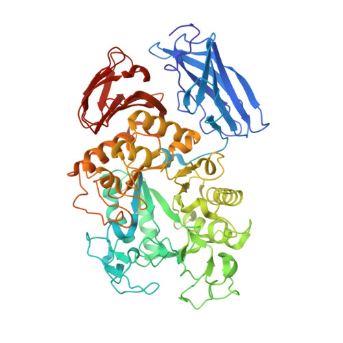
    Thermoactinomyces vulgaris R-47 alpha-amylase 1 (TVAI) has unique hydrolyzing activities for pullulan with sequence repeats of alpha-(1,4), alpha-(1,4), and alpha-(1,6) glycosidic linkages, as well as for starch. TVAI mainly hydrolyzes alpha-(1,4) glycosidic linkages to produce a panose, but it also hydrolyzes alpha-(1,6) glycosidic linkages with a lesser efficiency. X-ray structures of three complexes comprising an inactive mutant TVAI (D356N or D356N/E396Q) and a pullulan model oligosaccharide (P2; [Glc-alpha-(1,6)-Glc-alpha-(1,4)-Glc-alpha-(1,4)]2 or P5; [Glc-alpha-(1,6)-Glc-alpha-(1,4)-Glc-alpha-(1,4)]5) were determined. The complex D356N/P2 is a mimic of the enzyme/product complex in the main catalytic reaction of TVAI, and a structural comparison with Aspergillus oryzaealpha-amylase showed that the (-) subsites of TVAI are responsible for recognizing both starch and pullulan. D356N/E396Q/P2 and D356N/E396Q/P5 provided models of the enzyme/substrate complex recognizing the alpha-(1,6) glycosidic linkage at the hydrolyzing site. They showed that only subsites -1 and -2 at the nonreducing end of TVAI are effective in the hydrolysis of alpha-(1,6) glycosidic linkages, leading to weak interactions between substrates and the enzyme. Domain N of TVAI is a starch-binding domain acting as an anchor in the catalytic reaction of the enzyme. In this study, additional substrates were also found to bind to domain N, suggesting that domain N also functions as a pullulan-binding domain.


  • Organizational Affiliation

    Graduate School of Medicine, Kagawa University, Japan.


Macromolecules
Find similar proteins by:  (by identity cutoff)  |  3D Structure
Entity ID: 1
MoleculeChains Sequence LengthOrganismDetailsImage
alpha-amylase I637Thermoactinomyces vulgarisMutation(s): 2 
EC: 3.2.1.1
UniProt
Find proteins for Q60053 (Thermoactinomyces vulgaris)
Explore Q60053 
Go to UniProtKB:  Q60053
Entity Groups  
Sequence Clusters30% Identity50% Identity70% Identity90% Identity95% Identity100% Identity
UniProt GroupQ60053
Sequence Annotations
Expand
  • Reference Sequence
Oligosaccharides

Help

Entity ID: 2
MoleculeChains Length2D Diagram Glycosylation3D Interactions
beta-D-glucopyranose-(1-4)-alpha-D-glucopyranose-(1-6)-beta-D-glucopyranose-(1-4)-beta-D-glucopyranose-(1-4)-alpha-D-glucopyranose-(1-6)-alpha-D-glucopyranose-(1-4)-alpha-D-glucopyranose-(1-4)-alpha-D-glucopyranose-(1-6)-alpha-D-glucopyranose-(1-4)-alpha-D-glucopyranose
B
10N/A
Glycosylation Resources
GlyTouCan:  G39844GI
GlyCosmos:  G39844GI
Entity ID: 3
MoleculeChains Length2D Diagram Glycosylation3D Interactions
beta-D-glucopyranose-(1-4)-beta-D-glucopyranose
C
2N/A
Glycosylation Resources
GlyTouCan:  G84824ZO
GlyCosmos:  G84824ZO
Entity ID: 4
MoleculeChains Length2D Diagram Glycosylation3D Interactions
alpha-D-glucopyranose-(1-4)-beta-D-glucopyranose-(1-4)-beta-D-glucopyranose-(1-6)-beta-D-glucopyranose-(1-4)-beta-D-glucopyranose
D
5N/A
Glycosylation Resources
GlyTouCan:  G95107LS
GlyCosmos:  G95107LS
Biologically Interesting Molecules (External Reference) 1 Unique
Experimental Data & Validation

Experimental Data

  • Method: X-RAY DIFFRACTION
  • Resolution: 2.60 Å
  • R-Value Free: 0.190 
  • R-Value Work: 0.150 
  • R-Value Observed: 0.154 
  • Space Group: C 1 2 1
Unit Cell:
Length ( Å )Angle ( ˚ )
a = 120.885α = 90
b = 50.698β = 103.43
c = 108.007γ = 90
Software Package:
Software NamePurpose
MOSFLMdata reduction
SCALAdata scaling
CNSrefinement
CCP4data scaling
CNSphasing

Structure Validation

View Full Validation Report


Ligand Structure Quality Assessment 


Entry History 

Deposition Data

Revision History  (Full details and data files)

  • Version 1.0: 2006-07-11
    Type: Initial release
  • Version 1.1: 2008-04-30
    Changes: Version format compliance
  • Version 1.2: 2011-07-13
    Changes: Version format compliance
  • Version 2.0: 2020-07-29
    Type: Remediation
    Reason: Carbohydrate remediation
    Changes: Atomic model, Data collection, Derived calculations, Structure summary
  • Version 2.1: 2021-11-10
    Changes: Database references, Structure summary