2CH8

Structure of the Epstein-Barr Virus Oncogene BARF1


Experimental Data Snapshot

  • Method: X-RAY DIFFRACTION
  • Resolution: 2.30 Å
  • R-Value Free: 0.233 
  • R-Value Work: 0.174 
  • R-Value Observed: 0.177 

wwPDB Validation   3D Report Full Report


This is version 2.0 of the entry. See complete history


Literature

Structure of the Epstein-Barr Virus Oncogene Barf1

Tarbouriech, N.Ruggiero, F.Deturenne-Tessier, M.Ooka, T.Burmeister, W.P.

(2006) J Mol Biol 359: 667

  • DOI: https://doi.org/10.1016/j.jmb.2006.03.056
  • Primary Citation of Related Structures:  
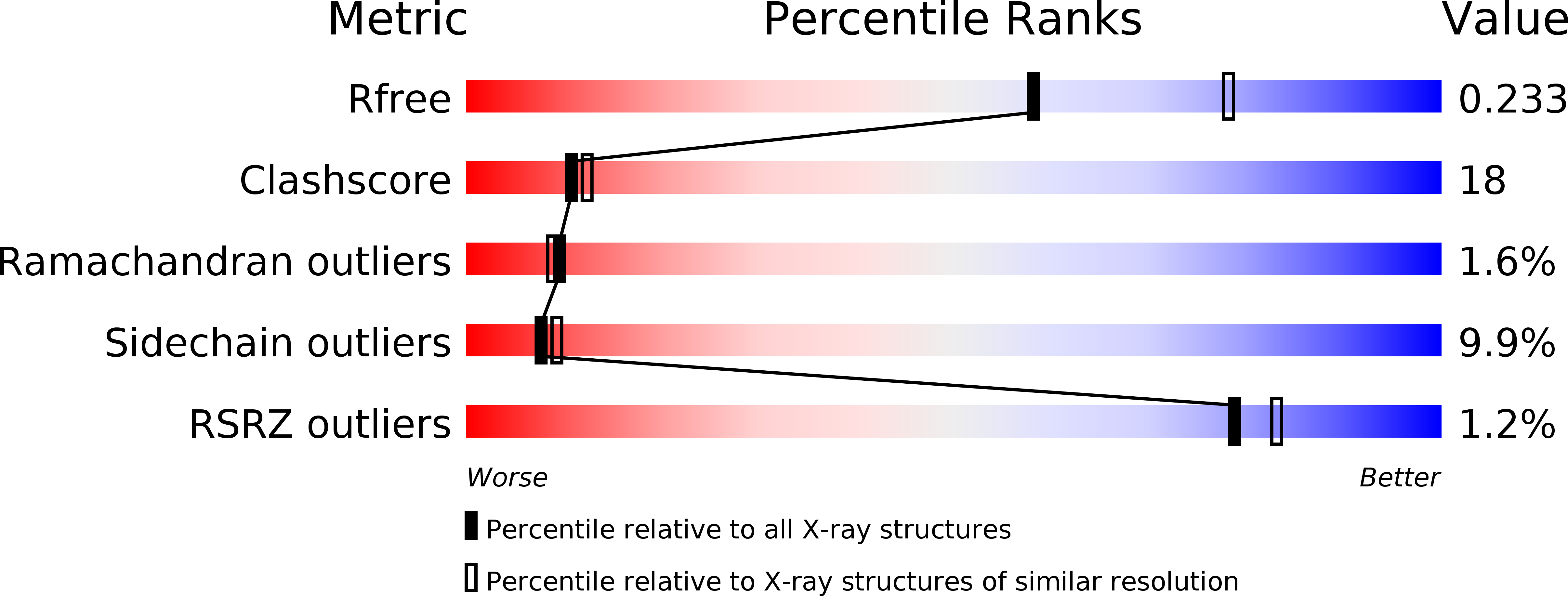
    2CH8

  • PubMed Abstract: 

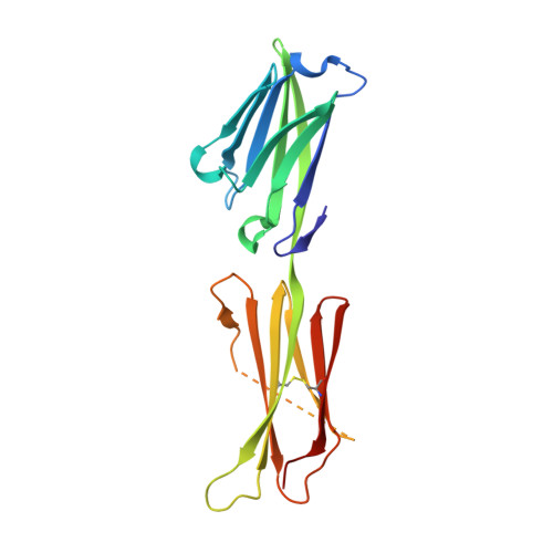
    The Epstein-Barr virus is a human gamma-herpesvirus that persistently infects more than 90% of the human population. It is associated with numerous epithelial cancers, principally undifferentiated nasopharyngeal carcinoma and gastric carcinoma. The BARF1 gene is expressed in a high proportion of these cancers. An oncogenic, mitogenic and immortalizing activity of the BARF1 protein has been shown. We solved the structure of the secreted BARF1 glycoprotein expressed in a human cell line by X-ray crystallography at a resolution of 2.3A. The BARF1 protein consists of two immunoglobulin (Ig)-like domains. The N-terminal domain belongs to the subfamily of variable domains whereas the C-terminal one is related to a constant Ig-domain. BARF1 shows an unusual hexamerisation involving two principal contacts, one between the C-terminal domains and one between the N-terminal domains. The C-terminal contact with an uncommonly large contact surface extends the beta-sandwich of the Ig-domain through the second molecule. The N-terminal contact involves Ig-domains with an unusual relative orientation but with a more classical contact surface with a size in the range of dimer interactions of Ig-domains. The structure of BARF1 is most closely related to CD80 or B7-1, a co-stimulatory molecule present on antigen presenting cells, from which BARF1 must have been derived during evolution. Still, domain orientation and oligomerization differ between BARF1 and CD80. It had been shown that BARF1 binds to hCSF-1, the human colony-stimulating factor 1, but this interaction has to be principally different from the one between CSF-1 and CSF-1 receptor.


  • Organizational Affiliation

    EMBL Grenoble outstation, BP181, F-38042, Grenoble cedex 9, France.


Macromolecules
Find similar proteins by:  (by identity cutoff)  |  3D Structure
Entity ID: 1
MoleculeChains Sequence LengthOrganismDetailsImage
33 KDA EARLY PROTEIN
A, B, C, D
201human gammaherpesvirus 4Mutation(s): 0 
UniProt
Find proteins for P03228 (Epstein-Barr virus (strain B95-8))
Explore P03228 
Go to UniProtKB:  P03228
Entity Groups  
Sequence Clusters30% Identity50% Identity70% Identity90% Identity95% Identity100% Identity
UniProt GroupP03228
Sequence Annotations
Expand
  • Reference Sequence
Oligosaccharides

Help

Entity ID: 2
MoleculeChains Length2D Diagram Glycosylation3D Interactions
alpha-D-mannopyranose-(1-4)-2-acetamido-2-deoxy-beta-D-glucopyranose-(1-4)-2-acetamido-2-deoxy-beta-D-glucopyranose
E, F, G, H
3N-Glycosylation
Glycosylation Resources
GlyTouCan:  G62182OO
GlyCosmos:  G62182OO
GlyGen:  G62182OO
Small Molecules
Ligands 1 Unique
IDChains Name / Formula / InChI Key2D Diagram3D Interactions
PT
Query on PT

Download Ideal Coordinates CCD File 
I [auth A]
J [auth A]
K [auth A]
L [auth B]
M [auth B]
I [auth A],
J [auth A],
K [auth A],
L [auth B],
M [auth B],
N [auth B],
O [auth C],
P [auth C],
Q [auth C],
R [auth D],
S [auth D],
T [auth D]
PLATINUM (II) ION
Pt
HRGDZIGMBDGFTC-UHFFFAOYSA-N
Experimental Data & Validation

Experimental Data

  • Method: X-RAY DIFFRACTION
  • Resolution: 2.30 Å
  • R-Value Free: 0.233 
  • R-Value Work: 0.174 
  • R-Value Observed: 0.177 
  • Space Group: H 3
Unit Cell:
Length ( Å )Angle ( ˚ )
a = 179.245α = 90
b = 179.245β = 90
c = 95.72γ = 120
Software Package:
Software NamePurpose
REFMACrefinement
MOSFLMdata reduction
SCALAdata scaling

Structure Validation

View Full Validation Report


Entry History 

Deposition Data

Revision History  (Full details and data files)

  • Version 1.0: 2006-05-31
    Type: Initial release
  • Version 1.1: 2011-07-13
    Changes: Advisory, Refinement description, Version format compliance
  • Version 2.0: 2020-07-29
    Type: Remediation
    Reason: Carbohydrate remediation
    Changes: Advisory, Atomic model, Data collection, Derived calculations, Other, Structure summary