2BX6

Crystal Structure of the human Retinitis Pigmentosa protein 2 (RP2)


Experimental Data Snapshot

  • Method: X-RAY DIFFRACTION
  • Resolution: 2.10 Å
  • R-Value Free: 0.235 
  • R-Value Work: 0.197 
  • R-Value Observed: 0.199 

wwPDB Validation   3D Report Full Report


This is version 1.1 of the entry. See complete history


Literature

Crystal Structure of the Human Retinitis Pigmentosa 2 Protein and its Interaction with Arl3

Kuhnel, K.Veltel, S.Schlichting, I.Wittinghofer, A.

(2006) Structure 14: 367

  • DOI: https://doi.org/10.1016/j.str.2005.11.008
  • Primary Citation of Related Structures:  
    2BX6

  • PubMed Abstract: 

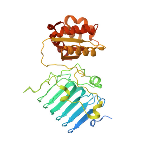
    The crystal structure of human retinitis pigmentosa 2 protein (RP2) was solved to 2.1 angstroms resolution. It consists of an N-terminal beta helix and a C-terminal ferredoxin-like alpha/beta domain. RP2 is functionally and structurally related to the tubulin-specific chaperone cofactor C. Seven of nine known RP2 missense mutations identified in patients are located in the beta helix domain, and most of them cluster to the hydrophobic core and are likely to destabilize the protein. Two residues, Glu138 and the catalytically important Arg118, are solvent-exposed and form a salt bridge, indicating that Glu138 might be critical for positioning Arg118 for catalysis. RP2 is a specific effector protein of Arl3. The N-terminal 34 residues and beta helix domain of RP2 are required for this interaction. The abilitities of RP2 to bind Arl3 and cause retinitis pigmentosa seem to be correlated, since both the R118H and E138G mutants show a drastically reduced affinity to Arl3.


  • Organizational Affiliation

    Max-Planck-Institut für Molekulare Physiologie, Abteilung Strukturelle Biologie, Otto-Hahn-Strasse 11, 44227 Dortmund, Germany.


Macromolecules
Find similar proteins by:  (by identity cutoff)  |  3D Structure
Entity ID: 1
MoleculeChains Sequence LengthOrganismDetailsImage
XRP2 PROTEIN350Homo sapiensMutation(s): 0 
UniProt & NIH Common Fund Data Resources
Find proteins for O75695 (Homo sapiens)
Explore O75695 
Go to UniProtKB:  O75695
PHAROS:  O75695
GTEx:  ENSG00000102218 
Entity Groups  
Sequence Clusters30% Identity50% Identity70% Identity90% Identity95% Identity100% Identity
UniProt GroupO75695
Sequence Annotations
Expand
  • Reference Sequence
Small Molecules
Ligands 1 Unique
IDChains Name / Formula / InChI Key2D Diagram3D Interactions
SO4
Query on SO4

Download Ideal Coordinates CCD File 
B [auth A]SULFATE ION
O4 S
QAOWNCQODCNURD-UHFFFAOYSA-L
Experimental Data & Validation

Experimental Data

  • Method: X-RAY DIFFRACTION
  • Resolution: 2.10 Å
  • R-Value Free: 0.235 
  • R-Value Work: 0.197 
  • R-Value Observed: 0.199 
  • Space Group: P 32 2 1
Unit Cell:
Length ( Å )Angle ( ˚ )
a = 81.3α = 90
b = 81.3β = 90
c = 106.12γ = 120
Software Package:
Software NamePurpose
REFMACrefinement
XDSdata reduction
XSCALEdata scaling
SHARPphasing

Structure Validation

View Full Validation Report


Entry History 

Deposition Data

Revision History  (Full details and data files)

  • Version 1.0: 2006-01-18
    Type: Initial release
  • Version 1.1: 2011-07-13
    Changes: Advisory, Version format compliance