2BTQ

Structure of BtubAB heterodimer from Prosthecobacter dejongeii


Experimental Data Snapshot

  • Method: X-RAY DIFFRACTION
  • Resolution: 3.20 Å
  • R-Value Free: 0.247 
  • R-Value Work: 0.215 
  • R-Value Observed: 0.215 

wwPDB Validation   3D Report Full Report


Ligand Structure Quality Assessment 


This is version 1.4 of the entry. See complete history


Literature

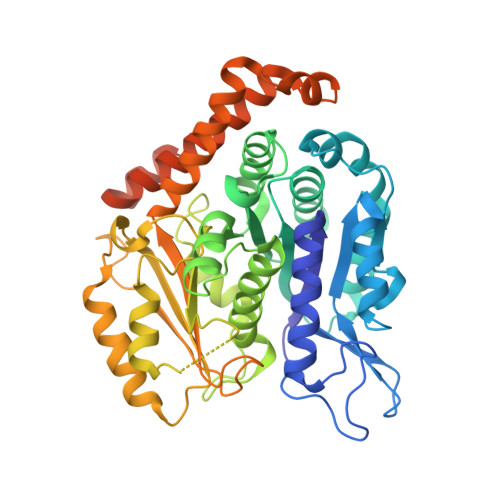
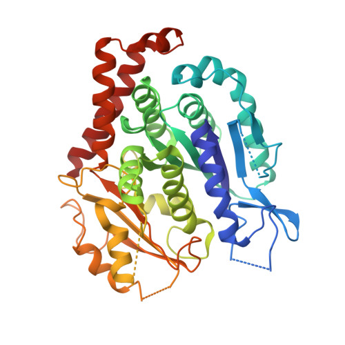
Structure of Bacterial Tubulin Btuba/B: Evidence for Horizontal Gene Transfer.

Schlieper, D.Oliva, M.A.Andreu, J.M.Lowe, J.

(2005) Proc Natl Acad Sci U S A 102: 9170

  • DOI: https://doi.org/10.1073/pnas.0502859102
  • Primary Citation of Related Structures:  
    2BTO, 2BTQ

  • PubMed Abstract: 

    alphabeta-Tubulin heterodimers, from which the microtubules of the cytoskeleton are built, have a complex chaperone-dependent folding pathway. They are thought to be unique to eukaryotes, whereas the homologue FtsZ can be found in bacteria. The exceptions are BtubA and BtubB from Prosthecobacter, which have higher sequence homology to eukaryotic tubulin than to FtsZ. Here we show that some of their properties are different from tubulin, such as weak dimerization and chaperone-independent folding. However, their structure is strikingly similar to tubulin including surface loops, and BtubA/B form tubulin-like protofilaments. Presumably, BtubA/B were transferred from a eukaryotic cell by horizontal gene transfer because their high degree of similarity to eukaryotic genes is unique within the Prosthecobacter genome.


  • Organizational Affiliation

    Laboratory of Molecular Biology, Medical Research Council, Hills Road, Cambridge CB2 2QH, United Kingdom.


Macromolecules
Find similar proteins by:  (by identity cutoff)  |  3D Structure
Entity ID: 1
MoleculeChains Sequence LengthOrganismDetailsImage
TUBULIN BTUBA473Prosthecobacter dejongeiiMutation(s): 0 
UniProt
Find proteins for Q8GCC5 (Prosthecobacter dejongeii)
Explore Q8GCC5 
Go to UniProtKB:  Q8GCC5
Entity Groups  
Sequence Clusters30% Identity50% Identity70% Identity90% Identity95% Identity100% Identity
UniProt GroupQ8GCC5
Sequence Annotations
Expand
  • Reference Sequence
Find similar proteins by:  (by identity cutoff)  |  3D Structure
Entity ID: 2
MoleculeChains Sequence LengthOrganismDetailsImage
TUBULIN BTUBB426Prosthecobacter dejongeiiMutation(s): 0 
UniProt
Find proteins for Q8GCC1 (Prosthecobacter dejongeii)
Explore Q8GCC1 
Go to UniProtKB:  Q8GCC1
Entity Groups  
Sequence Clusters30% Identity50% Identity70% Identity90% Identity95% Identity100% Identity
UniProt GroupQ8GCC1
Sequence Annotations
Expand
  • Reference Sequence
Experimental Data & Validation

Experimental Data

  • Method: X-RAY DIFFRACTION
  • Resolution: 3.20 Å
  • R-Value Free: 0.247 
  • R-Value Work: 0.215 
  • R-Value Observed: 0.215 
  • Space Group: P 65 2 2
Unit Cell:
Length ( Å )Angle ( ˚ )
a = 154.398α = 90
b = 154.398β = 90
c = 256.33γ = 120
Software Package:
Software NamePurpose
CNSrefinement
MOSFLMdata reduction
SCALAdata scaling

Structure Validation

View Full Validation Report


Ligand Structure Quality Assessment 


Entry History 

Deposition Data

Revision History  (Full details and data files)

  • Version 1.0: 2005-06-14
    Type: Initial release
  • Version 1.1: 2011-05-08
    Changes: Version format compliance
  • Version 1.2: 2011-07-13
    Changes: Version format compliance
  • Version 1.3: 2018-01-24
    Changes: Source and taxonomy
  • Version 1.4: 2023-12-13
    Changes: Data collection, Database references, Other, Refinement description