Crystal Structure of the P2 C-Repressor: A Binder of Non-Palindromic Direct DNA Repeats.
Massad, T., Skaar, K., Nilsson, H., Damberg, P., Henriksson-Peltola, P., Haggard-Ljungquist, E., Hogbom, M., Stenmark, P.(2010) Nucleic Acids Res 38: 7778
- PubMed: 20639540
- DOI: https://doi.org/10.1093/nar/gkq626
- Primary Citation of Related Structures:
2XCJ - PubMed Abstract:
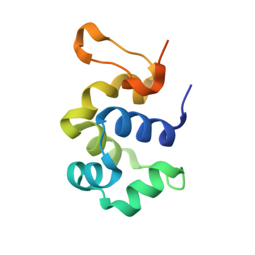
As opposed to the vast majority of prokaryotic repressors, the immunity repressor of temperate Escherichia coli phage P2 (C) recognizes non-palindromic direct repeats of DNA rather than inverted repeats. We have determined the crystal structure of P2 C at 1.8 Å. This constitutes the first structure solved from the family of C proteins from P2-like bacteriophages. The structure reveals that the P2 C protein forms a symmetric dimer oriented to bind the major groove of two consecutive turns of the DNA. Surprisingly, P2 C has great similarities to binders of palindromic sequences. Nevertheless, the two identical DNA-binding helixes of the symmetric P2 C dimer have to bind different DNA sequences. Helix 3 is identified as the DNA-recognition motif in P2 C by alanine scanning and the importance for the individual residues in DNA recognition is defined. A truncation mutant shows that the disordered C-terminus is dispensable for repressor function. The short distance between the DNA-binding helices together with a possible interaction between two P2 C dimers are proposed to be responsible for extensive bending of the DNA. The structure provides insight into the mechanisms behind the mutants of P2 C causing dimer disruption, temperature sensitivity and insensitivity to the P4 antirepressor.
Organizational Affiliation:
Department of Biochemistry and Biophysics, Center for Biomembrane Research, Stockholm University, SE-106 91 Stockholm, Sweden.