2RDV

RUBREDOXIN FROM DESULFOVIBRIO VULGARIS MIYAZAKI F, MONOCLINIC CRYSTAL FORM


Experimental Data Snapshot

  • Method: X-RAY DIFFRACTION
  • Resolution: 1.90 Å
  • R-Value Free: 0.216 
  • R-Value Work: 0.175 
  • R-Value Observed: 0.175 

wwPDB Validation   3D Report Full Report


This is version 1.3 of the entry. See complete history


Literature

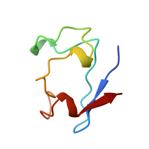
Structure determination of rubredoxin from Desulfovibrio vulgaris Miyazaki F in two crystal forms.

Misaki, S.Morimoto, Y.Ogata, M.Yagi, T.Higuchi, Y.Yasuoka, N.

(1999) Acta Crystallogr D Biol Crystallogr 55: 408-413

  • DOI: https://doi.org/10.1107/s0907444998011810
  • Primary Citation of Related Structures:  
    1RDV, 2RDV

  • PubMed Abstract: 

    The structures of two crystal forms (form I, P3221, a = b = 43.7, c = 50.7 A; form II, P21, a = 27.3, b = 44.9, c = 51.2 A and beta = 90. 6 degrees ) of the rubredoxin from Desulfovibrio vulgaris Miyazaki F have been solved by the molecular-replacement method. Form I has been refined at a resolution of 2.0 A to an R value of 20.8% and includes 32 water molecules. Form II includes 86 water molecules and has been refined at 1.9 A resolution to an R value of 17.5%. In form II, there are three molecules in the asymmetric unit with the molecules related by a non-crystallographic 32 symmetry axis. In both crystal forms, it was found that only a few residues effectively participate in the formation of intermolecular contacts along both the crystallographic (form I) and the non-crystallographic (form II) 32 axes. The crystal structure of the form II crystal is compared with those of other rubredoxin molecules from anaerobic bacteria. From this comparison, a similarity in the core region, which is composed of aromatic residues and includes the active centre, has been revealed.


  • Organizational Affiliation

    Department of Life Science, Faculty of Science, Himeji Institute of Technology, Kanaji 1475-2, Kamigori, Ako-gun, Hyogo 678-1297, Japan.


Macromolecules
Find similar proteins by:  (by identity cutoff)  |  3D Structure
Entity ID: 1
MoleculeChains Sequence LengthOrganismDetailsImage
RUBREDOXIN
A, B, C
52Nitratidesulfovibrio vulgaris str. 'Miyazaki FMutation(s): 0 
UniProt
Find proteins for P15412 (Desulfovibrio vulgaris (strain DSM 19637 / Miyazaki F))
Explore P15412 
Go to UniProtKB:  P15412
Entity Groups  
Sequence Clusters30% Identity50% Identity70% Identity90% Identity95% Identity100% Identity
UniProt GroupP15412
Sequence Annotations
Expand
  • Reference Sequence
Experimental Data & Validation

Experimental Data

  • Method: X-RAY DIFFRACTION
  • Resolution: 1.90 Å
  • R-Value Free: 0.216 
  • R-Value Work: 0.175 
  • R-Value Observed: 0.175 
  • Space Group: P 1 21 1
Unit Cell:
Length ( Å )Angle ( ˚ )
a = 27.3α = 90
b = 44.9β = 90.6
c = 51.2γ = 90
Software Package:
Software NamePurpose
X-PLORmodel building
X-PLORrefinement
WEISdata reduction
FSCALEdata scaling
X-PLORphasing

Structure Validation

View Full Validation Report


Entry History 

Deposition Data

Revision History  (Full details and data files)

  • Version 1.0: 1999-05-18
    Type: Initial release
  • Version 1.1: 2008-03-25
    Changes: Version format compliance
  • Version 1.2: 2011-07-13
    Changes: Version format compliance
  • Version 1.3: 2023-08-09
    Changes: Database references, Derived calculations, Other, Refinement description