2KGB

NMR solution of the regulatory domain cardiac F77W-Troponin C in complex with the cardiac Troponin I 144-163 switch peptide


Experimental Data Snapshot

  • Method: SOLUTION NMR
  • Conformers Calculated: 100 
  • Conformers Submitted: 20 
  • Selection Criteria: target function 

wwPDB Validation   3D Report Full Report


This is version 1.2 of the entry. See complete history


Literature

The effect of the cosolvent trifluoroethanol on a tryptophan side chain orientation in the hydrophobic core of troponin C.

Julien, O.Mercier, P.Crane, M.L.Sykes, B.D.

(2009) Protein Sci 18: 1165-1174

  • DOI: https://doi.org/10.1002/pro.121
  • Primary Citation of Related Structures:  
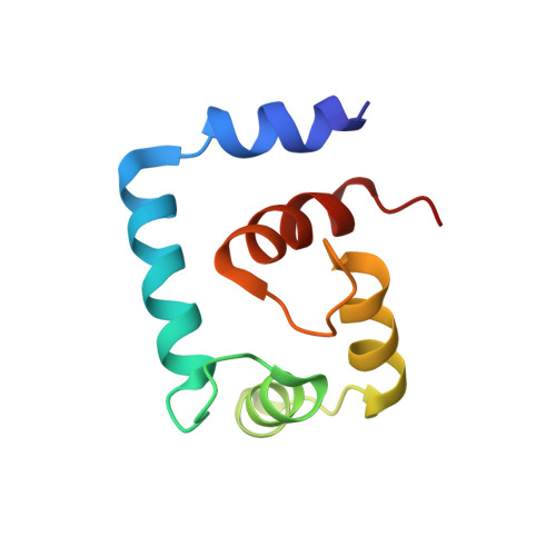
    2KGB

  • PubMed Abstract: 

    The unique biophysical properties of tryptophan residues have been exploited for decades to monitor protein structure and dynamics using a variety of spectroscopic techniques, such as fluorescence and nuclear magnetic resonance (NMR). We recently designed a tryptophan mutant in the regulatory N-domain of cardiac troponin C (F77W-cNTnC) to study the domain orientation of troponin C in muscle fibers using solid-state NMR. In our previous study, we determined the NMR structure of calcium-saturated mutant F77W-V82A-cNTnC in the presence of 19% 2,2,2-trifluoroethanol (TFE). TFE is a widely used cosolvent in the biophysical characterization of the solution structures of peptides and proteins. It is generally assumed that the structures are unchanged in the presence of cosolvents at relatively low concentrations, and this has been verified for TFE at the level of the overall secondary and tertiary structure for several calcium regulatory proteins. Here, we present the NMR solution structure of the calcium saturated F77W-cNTnC in presence of its biological binding partner troponin I peptide (cTnI(144-163)) and in the absence of TFE. We have also characterized a panel of six F77W-cNTnC structures in the presence and absence TFE, cTnI(144-163), and the extra mutation V82A, and used (19)F NMR to characterize the effect of TFE on the F77(5fW) analog. Our results show that although TFE did not perturb the overall protein structure, TFE did induce a change in the orientation of the indole ring of the buried tryptophan side chain from the anticipated position based upon homology with other proteins, highlighting the potential dangers of the use of cosolvents.


  • Organizational Affiliation

    Department of Biochemistry, University of Alberta, Edmonton, Alberta, Canada.


Macromolecules
Find similar proteins by:  (by identity cutoff)  |  3D Structure
Entity ID: 1
MoleculeChains Sequence LengthOrganismDetailsImage
Troponin C, slow skeletal and cardiac musclesA [auth C]89Homo sapiensMutation(s): 1 
Gene Names: TNNC1TNNC
UniProt & NIH Common Fund Data Resources
Find proteins for P63316 (Homo sapiens)
Explore P63316 
Go to UniProtKB:  P63316
PHAROS:  P63316
GTEx:  ENSG00000114854 
Entity Groups  
Sequence Clusters30% Identity50% Identity70% Identity90% Identity95% Identity100% Identity
UniProt GroupP63316
Sequence Annotations
Expand
  • Reference Sequence

Find similar proteins by:  Sequence   |   3D Structure  

Entity ID: 2
MoleculeChains Sequence LengthOrganismDetailsImage
Troponin I, cardiac muscleB [auth I]20Homo sapiensMutation(s): 0 
Gene Names: TNNI3
UniProt & NIH Common Fund Data Resources
Find proteins for P19429 (Homo sapiens)
Explore P19429 
Go to UniProtKB:  P19429
PHAROS:  P19429
GTEx:  ENSG00000129991 
Entity Groups  
Sequence Clusters30% Identity50% Identity70% Identity90% Identity95% Identity100% Identity
UniProt GroupP19429
Sequence Annotations
Expand
  • Reference Sequence
Small Molecules
Ligands 1 Unique
IDChains Name / Formula / InChI Key2D Diagram3D Interactions
CA
Query on CA

Download Ideal Coordinates CCD File 
C
CALCIUM ION
Ca
BHPQYMZQTOCNFJ-UHFFFAOYSA-N
Experimental Data & Validation

Experimental Data

  • Method: SOLUTION NMR
  • Conformers Calculated: 100 
  • Conformers Submitted: 20 
  • Selection Criteria: target function 

Structure Validation

View Full Validation Report


Entry History 

Deposition Data

Revision History  (Full details and data files)

  • Version 1.0: 2009-03-24
    Type: Initial release
  • Version 1.1: 2011-07-13
    Changes: Version format compliance
  • Version 1.2: 2021-10-20
    Changes: Database references, Derived calculations