2BKM

Crystal structure of the truncated hemoglobin from Geobacillus stearothermophilus


Experimental Data Snapshot

  • Method: X-RAY DIFFRACTION
  • Resolution: 1.50 Å
  • R-Value Free: 0.194 
  • R-Value Work: 0.167 
  • R-Value Observed: 0.168 

wwPDB Validation   3D Report Full Report


Ligand Structure Quality Assessment 


This is version 1.5 of the entry. See complete history


Literature

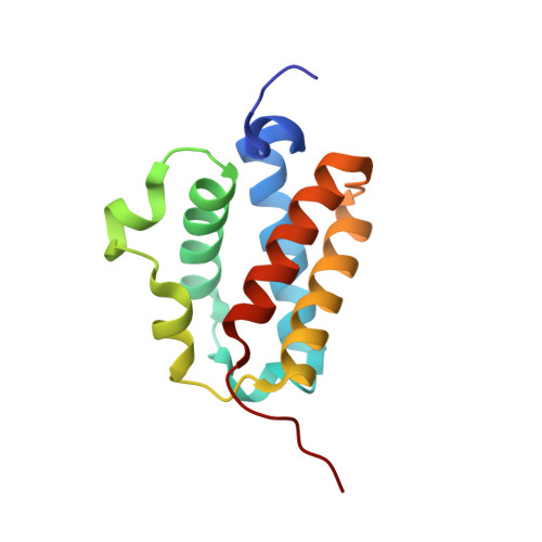
Crystal Structure and Ligand Binding Properties of the Truncated Hemoglobin from Geobacillus Stearothermophilus

Ilari, A.Kjelgaard, P.von Wachenfeldt, C.Catacchio, B.Chiancone, E.Boffi, A.

(2007) Arch Biochem Biophys 457: 85

  • DOI: https://doi.org/10.1016/j.abb.2006.09.033
  • Primary Citation of Related Structures:  
    2BKM

  • PubMed Abstract: 

    A novel truncated hemoglobin has been identified in the thermophilic bacterium Geobacillus stearothermophilus (Gs-trHb). The protein has been expressed in Escherichia coli, the 3D crystal structure (at 1.5 Angstroms resolution) and the ligand binding properties have been determined. The distal heme pocket displays an array of hydrogen bonding donors to the iron-bound ligands, including Tyr-B10 on one side of the heme pocket and Trp-G8 indole nitrogen on the opposite side. At variance with the highly similar Bacillus subtilis hemoglobin, Gs-trHb is dimeric both in the crystal and in solution and displays several unique structural properties. In the crystal cell, the iron-bound ligand is not homogeneously distributed within each distal site such that oxygen and an acetate anion can be resolved with relative occupancies of 50% each. Accordingly, equilibrium titrations of the oxygenated derivative in solution with acetate anion yield a partially saturated ferric acetate adduct. Moreover, the asymmetric unit contains two subunits and sedimentation velocity ultracentrifugation data confirm that the protein is dimeric.


  • Organizational Affiliation

    Department of Biochemical Sciences and CNR Institute of Molecular Biology and Pathology, University of Rome La Sapienza, P le Aldo Moro 5, 00185 Rome, Italy.


Macromolecules
Find similar proteins by:  (by identity cutoff)  |  3D Structure
Entity ID: 1
MoleculeChains Sequence LengthOrganismDetailsImage
TRUNCATED HEMOGLOBIN FROM GEOBACILLUS STEAROTHERMOPHILUS
A, B
128Geobacillus stearothermophilusMutation(s): 0 
UniProt
Find proteins for Q5L1S0 (Geobacillus kaustophilus (strain HTA426))
Explore Q5L1S0 
Go to UniProtKB:  Q5L1S0
Entity Groups  
Sequence Clusters30% Identity50% Identity70% Identity90% Identity95% Identity100% Identity
UniProt GroupQ5L1S0
Sequence Annotations
Expand
  • Reference Sequence
Experimental Data & Validation

Experimental Data

  • Method: X-RAY DIFFRACTION
  • Resolution: 1.50 Å
  • R-Value Free: 0.194 
  • R-Value Work: 0.167 
  • R-Value Observed: 0.168 
  • Space Group: P 1 21 1
Unit Cell:
Length ( Å )Angle ( ˚ )
a = 35.123α = 90
b = 99.339β = 105.59
c = 35.252γ = 90
Software Package:
Software NamePurpose
REFMACrefinement
DENZOdata reduction
SCALEPACKdata scaling
MOLREPphasing

Structure Validation

View Full Validation Report


Ligand Structure Quality Assessment 


Entry History 

Deposition Data

Revision History  (Full details and data files)

  • Version 1.0: 2006-11-29
    Type: Initial release
  • Version 1.1: 2011-05-08
    Changes: Version format compliance
  • Version 1.2: 2011-07-13
    Changes: Version format compliance
  • Version 1.3: 2017-07-12
    Changes: Derived calculations
  • Version 1.4: 2017-12-20
    Changes: Database references, Source and taxonomy, Structure summary
  • Version 1.5: 2023-12-13
    Changes: Data collection, Database references, Derived calculations, Other, Refinement description