2AI3

Purine nucleoside phosphorylase from calf spleen


Experimental Data Snapshot

  • Method: X-RAY DIFFRACTION
  • Resolution: 1.70 Å
  • R-Value Free: 0.253 
  • R-Value Work: 0.221 
  • R-Value Observed: 0.221 

wwPDB Validation   3D Report Full Report


Ligand Structure Quality Assessment 


This is version 1.3 of the entry. See complete history


Literature

Novel multisubstrate inhibitors of mammalian purine nucleoside phosphorylase.

Toms, A.V.Wang, W.Li, Y.Ganem, B.Ealick, S.E.

(2005) Acta Crystallogr D Biol Crystallogr 61: 1449-1458

  • DOI: https://doi.org/10.1107/S0907444905025503
  • Primary Citation of Related Structures:  
    2AI1, 2AI2, 2AI3

  • PubMed Abstract: 

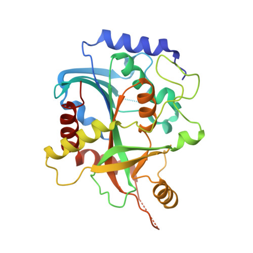
    In an effort to develop potent multisubstrate-analog inhibitors of purine nucleoside phosphorylase (PNP), three nucleoside phosphonates were designed utilizing structural information from the previously reported structures of complexes of bovine PNP with substrates and products. The nucleoside phosphonates contain an acetal linkage at the O2' and O3' positions and a two-C-atom spacer between the ribose and phosphate moieties. The linkage enables the compounds to simultaneously occupy the purine-, ribose- and phosphate-binding sites. The chemical syntheses, inhibition profiles and structural characterization of these novel multisubstrate analog inhibitors with bovine PNP are described.


  • Organizational Affiliation

    Department of Chemistry and Chemical Biology, Cornell University, Ithaca, NY 14853, USA.


Macromolecules
Find similar proteins by:  (by identity cutoff)  |  3D Structure
Entity ID: 1
MoleculeChains Sequence LengthOrganismDetailsImage
Purine nucleoside phosphorylase289Bos taurusMutation(s): 0 
EC: 2.4.2.1
UniProt
Find proteins for P55859 (Bos taurus)
Explore P55859 
Go to UniProtKB:  P55859
Entity Groups  
Sequence Clusters30% Identity50% Identity70% Identity90% Identity95% Identity100% Identity
UniProt GroupP55859
Sequence Annotations
Expand
  • Reference Sequence
Small Molecules
Ligands 4 Unique
IDChains Name / Formula / InChI Key2D Diagram3D Interactions
P2G
Query on P2G

Download Ideal Coordinates CCD File 
E [auth A](2S,4R,6R,6AS)-4-(2-AMINO-6-OXO-1,6-DIHYDROPURIN-9-YL)-6-(HYDROXYMETHYL)-TETRAHYDROFURO[3,4-D][1,3]DIOXOL-2-YLPHOSPHONI C ACID
C11 H14 N5 O8 P
GKAPYWCOOQBBHV-KXSYMAMXSA-N
144
Query on 144

Download Ideal Coordinates CCD File 
D [auth A]TRIS-HYDROXYMETHYL-METHYL-AMMONIUM
C4 H12 N O3
DRDCQJADRSJFFD-UHFFFAOYSA-N
ZN
Query on ZN

Download Ideal Coordinates CCD File 
C [auth A]ZINC ION
Zn
PTFCDOFLOPIGGS-UHFFFAOYSA-N
MG
Query on MG

Download Ideal Coordinates CCD File 
B [auth A]MAGNESIUM ION
Mg
JLVVSXFLKOJNIY-UHFFFAOYSA-N
Binding Affinity Annotations 
IDSourceBinding Affinity
P2G Binding MOAD:  2AI3 IC50: 5000 (nM) from 1 assay(s)
Experimental Data & Validation

Experimental Data

  • Method: X-RAY DIFFRACTION
  • Resolution: 1.70 Å
  • R-Value Free: 0.253 
  • R-Value Work: 0.221 
  • R-Value Observed: 0.221 
  • Space Group: P 21 3
Unit Cell:
Length ( Å )Angle ( ˚ )
a = 93.992α = 90
b = 93.992β = 90
c = 93.992γ = 90
Software Package:
Software NamePurpose
CNSrefinement
ADSCdata collection
HKL-2000data scaling
CNSphasing

Structure Validation

View Full Validation Report


Ligand Structure Quality Assessment 


Entry History 

Deposition Data

Revision History  (Full details and data files)

  • Version 1.0: 2005-10-25
    Type: Initial release
  • Version 1.1: 2008-04-30
    Changes: Version format compliance
  • Version 1.2: 2011-07-13
    Changes: Derived calculations, Version format compliance
  • Version 1.3: 2023-08-23
    Changes: Data collection, Database references, Derived calculations, Refinement description, Structure summary