Semi-extended Solution Structure of Human Myeloma Immunoglobulin D Determined by Constrained X-ray Scattering.
Sun, Z., Almogren, A., Furtado, P.B., Chowdhury, B., Kerr, M.A., Perkins, S.J.(2005) J Mol Biol 353: 155-173
- PubMed: 16157351
- DOI: https://doi.org/10.1016/j.jmb.2005.07.072
- Primary Citation of Related Structures:
1ZVO - PubMed Abstract:
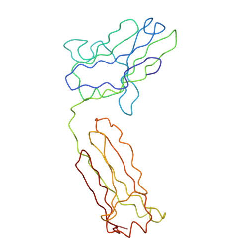
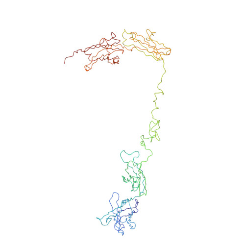
Human immunoglobulin D (IgD) occurs most abundantly as a membrane-bound antibody on the surface of mature B cells (mIgD). IgD possesses the longest hinge sequence of all the human antibody isotypes, with 64 residues connecting the Fab and Fc fragments. A novel rapid purification scheme of secreted IgD from the serum of an IgD myeloma patient using thiophilic (T-gel) and lectin affinity chromatography gave a stable, homogeneous IgD preparation. Synchrotron X-ray scattering and analytical ultracentrifugation of IgD identified the solution arrangement of its Fab and Fc fragments, and thereby its hinge structure. The Guinier X-ray radius of gyration R(G) of 6.9(+/-0.1)nm showed that IgD is more extended in solution than the immunoglobulin subclass IgA1 (R(G) of 6.1-6.2nm). Its distance distribution function P(r) showed a single peak at 4.7nm and a maximum dimension of 23nm. Velocity experiments gave a sedimentation coefficient of 6.3S, which is similar to that for IgA1 at 6.2S. The complete IgD structure was modelled using molecular dynamics to generate IgD hinge structures, to which homology models for the Fab and Fc fragments were connected. Good scattering curve fits were obtained with 18 semi-extended best fit IgD models that were filtered from 8500 trial models. These best-fit models showed that the IgD hinge does not correspond to an extended polypeptide structure. The averaged solution structure arrangement of the Fab and Fc fragments in IgD is principally T-shaped and flexible, with contribution from Y-shaped and inverted Y-shaped structures. Although the linear sequence of the IgD hinge is much longer, comparison with previous scattering modelling of IgA1 and IgA2(m)1 suggests that the hinge of IgA1 and IgD are more similar than might have been expected, Both possess flexible T-shaped solution structures, probably reflecting the presence of restraining O-linked sugars.
Organizational Affiliation:
Department of Biochemistry and Molecular Biology, Royal Free and University College Medical School, University College London, Gower Street, London WC1E 6BT, UK.