A True Autoactivating Enzyme: Structural insight into mannose-binding lectin-associated serine protease-2 activations
Gal, P., Harmat, V., Kocsis, A., Bian, T., Barna, L., Ambrus, G., Vegh, B., Balczer, J., Sim, R.B., Naray-Szabo, G., Zavodszky, P.(2005) J Biol Chem 280: 33435-33444
- PubMed: 16040602
- DOI: https://doi.org/10.1074/jbc.M506051200
- Primary Citation of Related Structures:
1ZJK - PubMed Abstract:
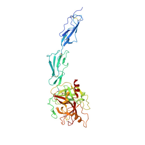
Few reports have described in detail a true autoactivation process, where no extrinsic cleavage factors are required to initiate the autoactivation of a zymogen. Herein, we provide structural and mechanistic insight into the autoactivation of a multidomain serine protease: mannose-binding lectin-associated serine protease-2 (MASP-2), the first enzymatic component in the lectin pathway of complement activation. We characterized the proenzyme form of a MASP-2 catalytic fragment encompassing its C-terminal three domains and solved its crystal structure at 2.4 A resolution. Surprisingly, zymogen MASP-2 is capable of cleaving its natural substrate C4, with an efficiency about 10% that of active MASP-2. Comparison of the zymogen and active structures of MASP-2 reveals that, in addition to the activation domain, other loops of the serine protease domain undergo significant conformational changes. This additional flexibility could play a key role in the transition of zymogen MASP-2 into a proteolytically active form. Based on the three-dimensional structures of proenzyme and active MASP-2 catalytic fragments, we present model for the active zymogen MASP-2 complex and propose a mechanism for the autoactivation process.
Organizational Affiliation:
Institute of Enzymology, Biological Research Center, Hungarian Academy of Sciences, P.O. Box 7, Budapest H-1518, Hungary.