1Z4O

Structure of beta-phosphoglucomutase with inhibitor bound alpha-galactose 1-phosphate


Experimental Data Snapshot

  • Method: X-RAY DIFFRACTION
  • Resolution: 1.90 Å
  • R-Value Free: 0.254 
  • R-Value Work: 0.199 
  • R-Value Observed: 0.205 

wwPDB Validation   3D Report Full Report


Ligand Structure Quality Assessment 


This is version 1.5 of the entry. See complete history


Literature

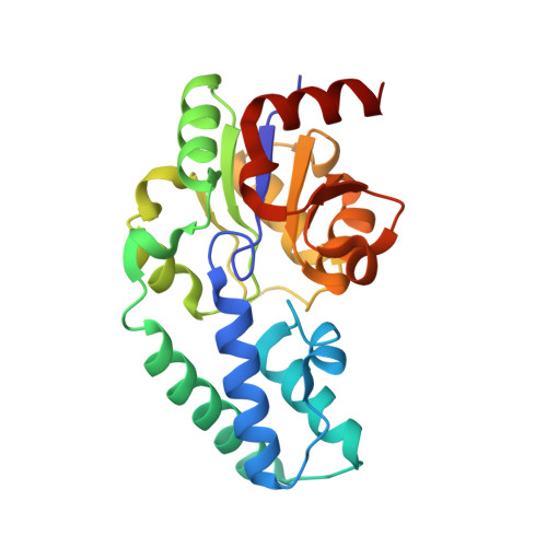
Chemical Confirmation of a Pentavalent Phosphorane in Complex with beta-Phosphoglucomutase

Tremblay, L.W.Zhang, G.Dai, J.Dunaway-Mariano, D.Allen, K.N.

(2005) J Am Chem Soc 127: 5298-5299

  • DOI: https://doi.org/10.1021/ja0509073
  • Primary Citation of Related Structures:  
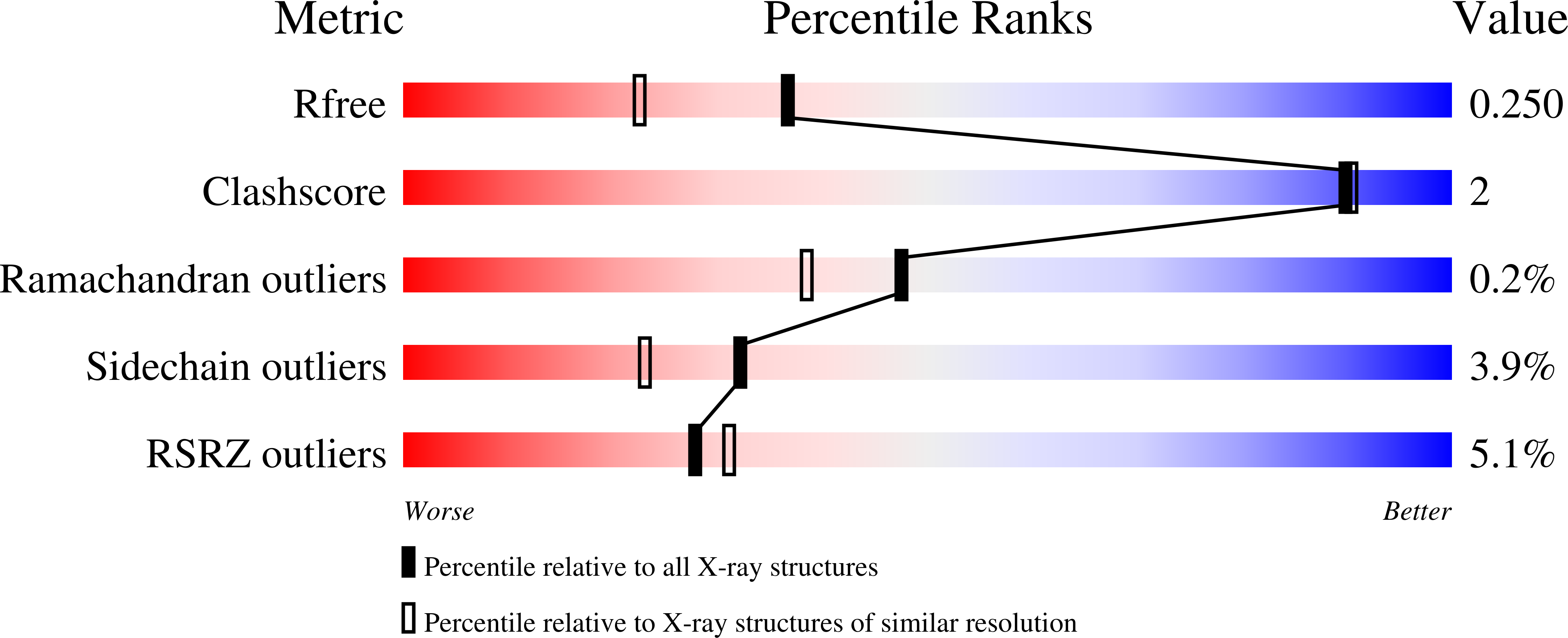
    1Z4N, 1Z4O

  • PubMed Abstract: 

    This communication reports the X-ray crystal structure of the alpha-d-galactose-1-phosphate complex with that of Lactococcus lactis beta-phosphoglucomutase (beta-PGM) crystallized in the presence of Mg2+ cofactor and the enzyme-to-phosphorus ratio determined by protein and phosphate analyses of the crystalline complex. The 1:1 ratio determined for this complex was compared to the 1:2 ratio determined for the crystals of beta-PGM grown in the presence of substrate and Mg2+ cofactor. This result verifies the published structure assignment of this latter complex as the phosphorane adduct formed by covalent bonding between the active site Asp8 carboxylate to the C(1)phosphorus of the beta-glucose 1,6-bisphosphate ligand and rules out the proposal of a beta-PGM-glucose-6-phosphate-1-MgF3- complex.


  • Organizational Affiliation

    Department of Physiology and Biophysics, Boston University School of Medicine, Boston, Massachusetts 02118-2394, USA.


Macromolecules
Find similar proteins by:  (by identity cutoff)  |  3D Structure
Entity ID: 1
MoleculeChains Sequence LengthOrganismDetailsImage
Beta-phosphoglucomutase
A, B
221Lactococcus lactisMutation(s): 0 
Gene Names: pgmB
EC: 5.4.2.6
UniProt
Find proteins for P71447 (Lactococcus lactis subsp. lactis (strain IL1403))
Explore P71447 
Go to UniProtKB:  P71447
Entity Groups  
Sequence Clusters30% Identity50% Identity70% Identity90% Identity95% Identity100% Identity
UniProt GroupP71447
Sequence Annotations
Expand
  • Reference Sequence
Small Molecules
Binding Affinity Annotations 
IDSourceBinding Affinity
GL1 PDBBind:  1Z4O Ki: 3.00e+4 (nM) from 1 assay(s)
Binding MOAD:  1Z4O Ki: 3.00e+4 (nM) from 1 assay(s)
Experimental Data & Validation

Experimental Data

  • Method: X-RAY DIFFRACTION
  • Resolution: 1.90 Å
  • R-Value Free: 0.254 
  • R-Value Work: 0.199 
  • R-Value Observed: 0.205 
  • Space Group: P 1 21 1
Unit Cell:
Length ( Å )Angle ( ˚ )
a = 55.069α = 90
b = 36.351β = 101.77
c = 104.883γ = 90
Software Package:
Software NamePurpose
DENZOdata reduction
SCALEPACKdata scaling
SHELXSphasing
CNSrefinement

Structure Validation

View Full Validation Report


Ligand Structure Quality Assessment 


Entry History 

Deposition Data

Revision History  (Full details and data files)

  • Version 1.0: 2005-04-19
    Type: Initial release
  • Version 1.1: 2008-04-30
    Changes: Version format compliance
  • Version 1.2: 2011-07-13
    Changes: Version format compliance
  • Version 1.3: 2018-01-31
    Changes: Experimental preparation
  • Version 1.4: 2020-07-29
    Type: Remediation
    Reason: Carbohydrate remediation
    Changes: Data collection, Derived calculations
  • Version 1.5: 2023-08-23
    Changes: Data collection, Database references, Refinement description, Structure summary