1XI1

Phi29 DNA polymerase ssDNA complex, monoclinic crystal form


Experimental Data Snapshot

  • Method: X-RAY DIFFRACTION
  • Resolution: 2.20 Å
  • R-Value Free: 0.277 
  • R-Value Work: 0.243 
  • R-Value Observed: 0.243 

wwPDB Validation   3D Report Full Report


This is version 1.5 of the entry. See complete history


Literature

Correction of X-ray intensities from single crystals containing lattice-translocation defects

Wang, J.Kamtekar, S.Berman, A.J.Steitz, T.A.

(2005) Acta Crystallogr D Biol Crystallogr 61: 67-74

  • DOI: https://doi.org/10.1107/S0907444904026721
  • Primary Citation of Related Structures:  
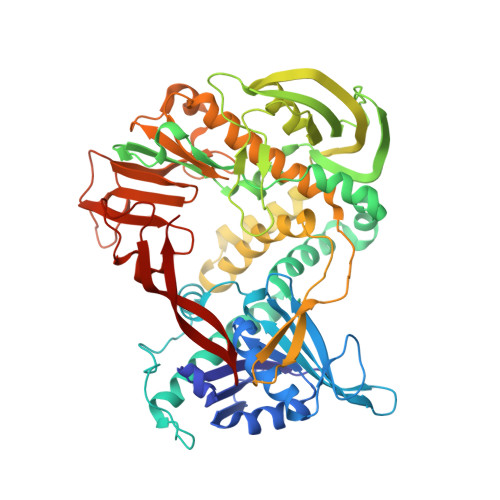
    1XI1

  • PubMed Abstract: 

    In 1954, Howells and colleagues described an unusual diffraction pattern from imidazole methemoglobin crystals caused by lattice-translocation defects. In these crystals, two identical lattices coexist as a single coherent mosaic block, but are translated by a fixed vector with respect to each other. The observed structure is a weighted sum of the two identical but translated structures, one from each lattice; the observed structure factors are a weighted vector sum of the two structure factors with identical unit amplitudes but shifted phases. A general procedure is described to obtain the unit amplitudes of observed structure factors from a realigned single lattice through an X-ray intensity correction. An application of this procedure is made to determine the crystal structure of phi29 DNA polymerase at 2.2 A resolution using multiple isomorphous replacement and multiwavelength anomalous dispersion methods.


  • Organizational Affiliation

    Department of Molecular Biophysics and Biochemistry, Yale University, 266 Whitney Avenue, New Haven, CT 06520-8114, USA. wang@mail.csb.yale.edu


Macromolecules

Find similar proteins by:  (by identity cutoff)  |  3D Structure
Entity ID: 2
MoleculeChains Sequence LengthOrganismDetailsImage
DNA polymeraseC [auth A],
D [auth B]
575Salasvirus phi29Mutation(s): 2 
Gene Names: 2
EC: 2.7.7.7
UniProt
Find proteins for P03680 (Bacillus phage phi29)
Explore P03680 
Go to UniProtKB:  P03680
Entity Groups  
Sequence Clusters30% Identity50% Identity70% Identity90% Identity95% Identity100% Identity
UniProt GroupP03680
Sequence Annotations
Expand
  • Reference Sequence

Find similar nucleic acids by:  Sequence   |   3D Structure  

Entity ID: 1
MoleculeChains LengthOrganismImage
5'-D(P*TP*TP*TP*TP*T)-3'A [auth C],
B [auth D]
5N/A
Sequence Annotations
Expand
  • Reference Sequence
Experimental Data & Validation

Experimental Data

  • Method: X-RAY DIFFRACTION
  • Resolution: 2.20 Å
  • R-Value Free: 0.277 
  • R-Value Work: 0.243 
  • R-Value Observed: 0.243 
  • Space Group: P 1 21 1
Unit Cell:
Length ( Å )Angle ( ˚ )
a = 60.557α = 90
b = 170.563β = 106.99
c = 68.813γ = 90
Software Package:
Software NamePurpose
CNSrefinement
HKL-2000data reduction
SCALEPACKdata scaling
MLPHAREphasing

Structure Validation

View Full Validation Report


Entry History 

Deposition Data

Revision History  (Full details and data files)

  • Version 1.0: 2004-12-07
    Type: Initial release
  • Version 1.1: 2008-04-30
    Changes: Version format compliance
  • Version 1.2: 2011-07-13
    Changes: Version format compliance
  • Version 1.3: 2020-01-22
    Changes: Advisory, Data collection, Derived calculations
  • Version 1.4: 2021-10-20
    Changes: Database references, Derived calculations
  • Version 1.5: 2024-02-14
    Changes: Data collection