1XE3

Crystal Structure of purine nucleoside phosphorylase DeoD from Bacillus anthracis


Experimental Data Snapshot

  • Method: X-RAY DIFFRACTION
  • Resolution: 2.24 Å
  • R-Value Free: 0.235 
  • R-Value Work: 0.181 
  • R-Value Observed: 0.184 

wwPDB Validation   3D Report Full Report


This is version 1.3 of the entry. See complete history


Literature

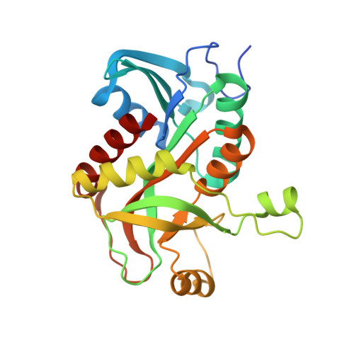
Structure of purine nucleoside phosphorylase (DeoD) from Bacillus anthracis.

Grenha, R.Levdikov, V.M.Fogg, M.J.Blagova, E.V.Brannigan, J.A.Wilkinson, A.J.Wilson, K.S.

(2005) Acta Crystallogr Sect F Struct Biol Cryst Commun 61: 459-462

  • DOI: https://doi.org/10.1107/S174430910501095X
  • Primary Citation of Related Structures:  
    1XE3

  • PubMed Abstract: 

    Protein structures from the causative agent of anthrax (Bacillus anthracis) are being determined as part of a structural genomics programme. Amongst initial candidates for crystallographic analysis are enzymes involved in nucleotide biosynthesis, since these are recognized as potential targets in antibacterial therapy. Purine nucleoside phosphorylase is a key enzyme in the purine-salvage pathway. The crystal structure of purine nucleoside phosphorylase (DeoD) from B. anthracis has been solved by molecular replacement at 2.24 A resolution and refined to an R factor of 18.4%. This is the first report of a DeoD structure from a Gram-positive bacterium.


  • Organizational Affiliation

    Department of Chemistry, University of York, York YO10 5YW, England.


Macromolecules
Find similar proteins by:  (by identity cutoff)  |  3D Structure
Entity ID: 1
MoleculeChains Sequence LengthOrganismDetailsImage
purine nucleoside phosphorylase
A, B, C, D, E
A, B, C, D, E, F
245Bacillus anthracisMutation(s): 0 
Gene Names: deoD
EC: 2.4.2.1
UniProt
Find proteins for Q81T09 (Bacillus anthracis)
Explore Q81T09 
Go to UniProtKB:  Q81T09
Entity Groups  
Sequence Clusters30% Identity50% Identity70% Identity90% Identity95% Identity100% Identity
UniProt GroupQ81T09
Sequence Annotations
Expand
  • Reference Sequence
Experimental Data & Validation

Experimental Data

  • Method: X-RAY DIFFRACTION
  • Resolution: 2.24 Å
  • R-Value Free: 0.235 
  • R-Value Work: 0.181 
  • R-Value Observed: 0.184 
  • Space Group: P 21 21 21
Unit Cell:
Length ( Å )Angle ( ˚ )
a = 63.861α = 90
b = 128.257β = 90
c = 223.565γ = 90
Software Package:
Software NamePurpose
REFMACrefinement
DENZOdata reduction
SCALEPACKdata scaling
MOLREPphasing

Structure Validation

View Full Validation Report


Entry History 

Revision History  (Full details and data files)

  • Version 1.0: 2004-10-26
    Type: Initial release
  • Version 1.1: 2008-04-30
    Changes: Version format compliance
  • Version 1.2: 2011-07-13
    Changes: Version format compliance
  • Version 1.3: 2023-08-23
    Changes: Data collection, Database references, Derived calculations, Refinement description