


Structural comparison of Nas6p protein structures in two different crystal forms
Nakamura, Y., Umehara, T., Tanaka, A., Horikoshi, M., Yokoyama, S., Padmanabhan, B.To be published.
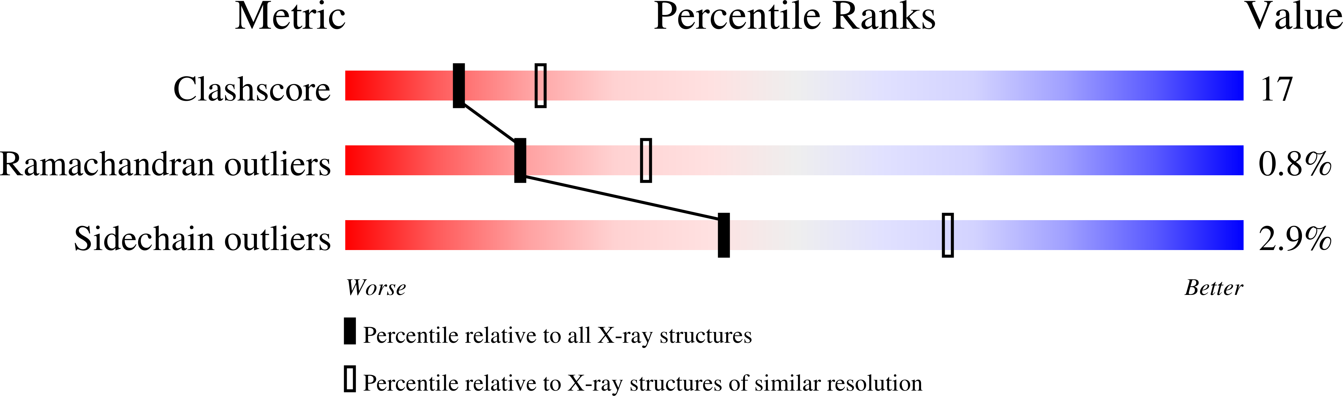
Experimental Data Snapshot
wwPDB Validation 3D Report Full Report
Entity ID: 1 | |||||
|---|---|---|---|---|---|
| Molecule | Chains | Sequence Length | Organism | Details | Image |
| Probable 26S proteasome regulatory subunit p28 | 243 | Saccharomyces cerevisiae | Mutation(s): 0 | 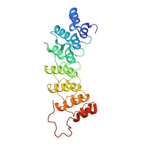 | |
UniProt | |||||
Find proteins for P50086 (Saccharomyces cerevisiae (strain ATCC 204508 / S288c)) Explore P50086 Go to UniProtKB: P50086 | |||||
Entity Groups | |||||
| Sequence Clusters | 30% Identity50% Identity70% Identity90% Identity95% Identity100% Identity | ||||
| UniProt Group | P50086 | ||||
Sequence AnnotationsExpand | |||||
| |||||
| Length ( Å ) | Angle ( ˚ ) |
|---|---|
| a = 43.444 | α = 90 |
| b = 68.629 | β = 108.95 |
| c = 46.985 | γ = 90 |
| Software Name | Purpose |
|---|---|
| HKL-2000 | data collection |
| SCALEPACK | data scaling |
| MOLREP | phasing |
| CNS | refinement |
| HKL-2000 | data reduction |
RCSB PDB (citation) is hosted by
RCSB PDB is a member of the

