1TGX

X-RAY STRUCTURE AT 1.55 A OF TOXIN GAMMA, A CARDIOTOXIN FROM NAJA NIGRICOLLIS VENOM. CRYSTAL PACKING REVEALS A MODEL FOR INSERTION INTO MEMBRANES


Experimental Data Snapshot

  • Method: X-RAY DIFFRACTION
  • Resolution: 1.55 Å
  • R-Value Work: 0.172 
  • R-Value Observed: 0.172 

wwPDB Validation   3D Report Full Report


This is version 1.5 of the entry. See complete history


Literature

X-ray structure at 1.55 A of toxin gamma, a cardiotoxin from Naja nigricollis venom. Crystal packing reveals a model for insertion into membranes.

Bilwes, A.Rees, B.Moras, D.Menez, R.Menez, A.

(1994) J Mol Biol 239: 122-136

  • DOI: https://doi.org/10.1006/jmbi.1994.1357
  • Primary Citation of Related Structures:  
    1TGX

  • PubMed Abstract: 

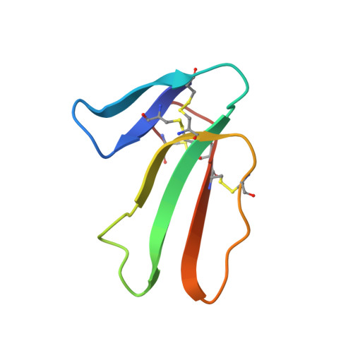
    The crystal structure of toxin gamma from Naja nigricollis has been solved and refined to 1.55 A resolution. The final R-factor, computed with all X-ray data available, is 17.9%. The three-dimensional structure is characterized by a core formed by two beta-sheets organized in three extended loops. It is similar to that of cardiotoxin V4II from Naja mossambica mossambica, with the exception of the hydrophobic loop I. The flexibility and variability of the loops contrast sharply with the rigidity of the molecular core and its high degree of structural conservation among the cardiotoxin family. The most flexible loop II adopts different conformations in the three monomers forming the crystal asymmetric unit. These monomers form a trimer around an approximate 3-fold axis, with conserved hydrophobic side-chains on the outside and hydrophilic residues in the central channel or involved in interactions with the other molecules. The trimer thus resembles a membrane protein with a central channel that could allow the passage of small ions. It is proposed as a model for the insertion of cardiotoxin into a membrane.


  • Organizational Affiliation

    UPR de Biologie Structurale, Institut de Biologie Moléculaire et Cellulaire du CNRS, Strasbourg, France.


Macromolecules
Find similar proteins by:  (by identity cutoff)  |  3D Structure
Entity ID: 1
MoleculeChains Sequence LengthOrganismDetailsImage
GAMMA-CARDIOTOXIN
A, B, C
60Naja nigricollisMutation(s): 0 
Membrane Entity: Yes 
UniProt
Find proteins for P01468 (Naja pallida)
Explore P01468 
Go to UniProtKB:  P01468
Entity Groups  
Sequence Clusters30% Identity50% Identity70% Identity90% Identity95% Identity100% Identity
UniProt GroupP01468
Sequence Annotations
Expand
  • Reference Sequence
Small Molecules
Ligands 1 Unique
IDChains Name / Formula / InChI Key2D Diagram3D Interactions
CL
Query on CL

Download Ideal Coordinates CCD File 
D [auth A]
E [auth A]
F [auth A]
G [auth A]
H [auth A]
D [auth A],
E [auth A],
F [auth A],
G [auth A],
H [auth A],
I [auth B],
J [auth B],
K [auth B],
L [auth B],
M [auth B],
N [auth B],
O [auth C],
P [auth C],
Q [auth C]
CHLORIDE ION
Cl
VEXZGXHMUGYJMC-UHFFFAOYSA-M
Experimental Data & Validation

Experimental Data

  • Method: X-RAY DIFFRACTION
  • Resolution: 1.55 Å
  • R-Value Work: 0.172 
  • R-Value Observed: 0.172 
  • Space Group: C 1 2 1
Unit Cell:
Length ( Å )Angle ( ˚ )
a = 78.67α = 90
b = 40.71β = 117.08
c = 56.02γ = 90
Software Package:
Software NamePurpose
X-PLORmodel building
PROLSQrefinement
X-PLORrefinement
X-PLORphasing

Structure Validation

View Full Validation Report


Entry History 

Deposition Data

Revision History  (Full details and data files)

  • Version 1.0: 1994-04-30
    Type: Initial release
  • Version 1.1: 2008-03-24
    Changes: Version format compliance
  • Version 1.2: 2011-07-13
    Changes: Version format compliance
  • Version 1.3: 2017-11-29
    Changes: Derived calculations, Other
  • Version 1.4: 2019-07-17
    Changes: Data collection, Refinement description
  • Version 1.5: 2019-08-14
    Changes: Data collection, Refinement description