Design of amino acid aldehydes as transition-state analogue inhibitors of arginase
Shin, H., Cama, E., Christianson, D.W.(2004) J Am Chem Soc 126: 10278-10284
- PubMed: 15315440
- DOI: https://doi.org/10.1021/ja047788w
- Primary Citation of Related Structures:
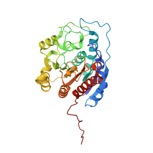
1T5F - PubMed Abstract:
Arginase is a binuclear manganese metalloenzyme that catalyzes the hydrolysis of l-arginine to form l-ornithine and urea. Chiral L-amino acids bearing aldehyde side chains have been synthesized in which the electrophilic aldehyde C=O bond is isosteric with the C=N bond of L-arginine. This substitution is intended to facilitate nucleophilic attack by the metal-bridging hydroxide ion upon binding to the arginase active site. Syntheses of the amino acid aldehydes have been accomplished by reduction, oxidation, and Wittig-type reaction with a commercially available derivative of L-glutamic acid. Amino acid aldehydes exhibit inhibition in the micromolar range, and the X-ray crystal structure of arginase I complexed with one of these inhibitors, (S)-2-amino-7-oxoheptanoic acid, has been determined at 2.2 A resolution. In the enzyme-inhibitor complex, the inhibitor aldehyde moiety is hydrated to form the gem-diol: one hydroxyl group bridges the Mn(2+)(2) cluster and donates a hydrogen bond to D128, and the second hydroxyl group donates a hydrogen bond to E277. The binding mode of the neutral gem-diol may mimic the binding of the neutral tetrahedral intermediate and its flanking transition states in arginase catalysis.
Organizational Affiliation:
Contribution from the Roy and Diana Vagelos Laboratories, Department of Chemistry, University of Pennsylvania, Philadelphia, PA 19104-6323, USA.