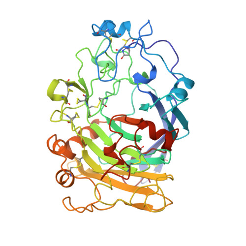
Structure of the Fusarium oxysporum endoglucanase I with a nonhydrolyzable substrate analogue: substrate distortion gives rise to the preferred axial orientation for the leaving group.
Sulzenbacher, G., Driguez, H., Henrissat, B., Schulein, M., Davies, G.J.(1996) Biochemistry 35: 15280-15287
- PubMed: 8952478
- DOI: https://doi.org/10.1021/bi961946h
- Primary Citation of Related Structures:
1OVW - PubMed Abstract:
Endoglucanase I (EG I) is a cellulase, from glycosyl hydrolase family 7, which cleaves the beta-1,4 linkages of cellulose with overall retention of configuration. The structure of the EG I from Fusarium oxysproum, complexed to a nonhydrolyzable thiooligosaccharide substrate analogue, has been determined by X-ray crystallography at a resolution of 2.7 A utilizing the 4-fold noncrystallographic symmetry present in the asymmetric unit. The electron density map clearly reveals the presence of three glucosyl units of the inhibitor, consistent with the known number of sugar-binding subsites, located at the active site of the enzyme in the -2, -1, and +1 subsites, i.e., actually spanning the point of enzymatic cleavage. The pyranose ring at the point of potential enzymatic cleavage is clearly distorted from the standard 4C1 chair as was originally suggested for beta-retaining enzymes by Phillips [Ford, L.O., Johnson, L.N., Machin, P. A., Phillips, D.C., & Tijan, T. (1974) J. Mol. Biol, 88, 349-371]. The distortion observed goes beyond the "sofa" conformation observed in previous studies and results in a conformation whose salient feature is the resulting quasi-axial orientation for the glycosidic bond and leaving group, as predicted by stereoelectronic theory. An almost identical conformation has recently been observed in a complex of chitobiase with its unhydrolyzed substrate [Tews, I., Perrakis, A., Oppenheim, A., Dauter, Z., Wilson, K. S., & Vorgias, C. E. (1996) Nat. Struct. Biol. 3, 638-648]. The striking similarity between these two complexes extends beyond the almost identical pyranose ring distortion. The overlap of the two respective sugars places the enzymatic nucleophile of endoglucanase I in coincidence with the C2 acetamido oxygen of N-acetylglucosamine in the catalytic site of the chitobiase, substantiating the involvement of this group in the catalytic mechanism of chitobiase and related chitinolytic enzymes. The endoglucanase I complex with the thiosaccharide substrate analogue clearly illustrates the potential of nonhydrolyzable sulfur-linked oligosaccharides in the elucidation of substrate binding and catalysis by glycosyl hydrolases.
Organizational Affiliation:
Department of Chemistry, University of York, Heslington, England.