1NPB

Crystal structure of the fosfomycin resistance protein from transposon Tn2921


Experimental Data Snapshot

  • Method: X-RAY DIFFRACTION
  • Resolution: 2.50 Å
  • R-Value Free: 0.230 
  • R-Value Work: 0.183 
  • R-Value Observed: 0.185 

wwPDB Validation   3D Report Full Report


This is version 1.3 of the entry. See complete history


Literature

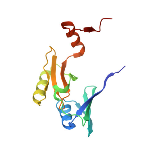
Structure of fosfomycin resistance protein FosA from transposon Tn2921.

Pakhomova, S.Rife, C.L.Armstrong, R.N.Newcomer, M.E.

(2004) Protein Sci 13: 1260-1265

  • DOI: https://doi.org/10.1110/ps.03585004
  • Primary Citation of Related Structures:  
    1NPB

  • PubMed Abstract: 

    The crystal structure of fosfomycin resistance protein FosA from transposon Tn2921 has been established at a resolution of 2.5 A. The protein crystallized without bound Mn(II) and K+, ions crucial for efficient catalysis, providing a structure of the apo enzyme. The protein maintains the three-dimensional domain-swapped arrangement of the paired betaalphabetabetabeta-motifs observed in the genomically encoded homologous enzyme from Pseudomonas aeruginosa (PA1129). The basic architecture of the active site is also maintained, despite the absence of the catalytically essential Mn(II). However, the absence of K+, which has been shown to enhance enzymatic activity, appears to contribute to conformational heterogeneity in the K(+)-binding loops.


  • Organizational Affiliation

    Departments of Biological Sciences and Chemistry, Louisiana State University, Baton Rouge, Louisiana 70803, USA. sveta@lsu.edu


Macromolecules
Find similar proteins by:  (by identity cutoff)  |  3D Structure
Entity ID: 1
MoleculeChains Sequence LengthOrganismDetailsImage
fosfomycin-resistance protein
A, B, C, D, E
A, B, C, D, E, F
141Serratia marcescensMutation(s): 0 
Gene Names: TN2921
UniProt
Find proteins for Q56415 (Serratia marcescens)
Explore Q56415 
Go to UniProtKB:  Q56415
Entity Groups  
Sequence Clusters30% Identity50% Identity70% Identity90% Identity95% Identity100% Identity
UniProt GroupQ56415
Sequence Annotations
Expand
  • Reference Sequence
Small Molecules
Ligands 2 Unique
IDChains Name / Formula / InChI Key2D Diagram3D Interactions
SO4
Query on SO4

Download Ideal Coordinates CCD File 
G [auth A]
H [auth A]
J [auth B]
L [auth C]
M [auth C]
G [auth A],
H [auth A],
J [auth B],
L [auth C],
M [auth C],
O [auth D],
P [auth D],
R [auth E],
S [auth E],
U [auth F]
SULFATE ION
O4 S
QAOWNCQODCNURD-UHFFFAOYSA-L
GOL
Query on GOL

Download Ideal Coordinates CCD File 
I [auth A]
K [auth B]
N [auth C]
Q [auth D]
T [auth E]
I [auth A],
K [auth B],
N [auth C],
Q [auth D],
T [auth E],
V [auth F],
W [auth F]
GLYCEROL
C3 H8 O3
PEDCQBHIVMGVHV-UHFFFAOYSA-N
Experimental Data & Validation

Experimental Data

  • Method: X-RAY DIFFRACTION
  • Resolution: 2.50 Å
  • R-Value Free: 0.230 
  • R-Value Work: 0.183 
  • R-Value Observed: 0.185 
  • Space Group: I 4 2 2
Unit Cell:
Length ( Å )Angle ( ˚ )
a = 208.597α = 90
b = 208.597β = 90
c = 136.358γ = 90
Software Package:
Software NamePurpose
REFMACrefinement
DENZOdata reduction
SCALEPACKdata scaling
AMoREphasing

Structure Validation

View Full Validation Report


Entry History 

Deposition Data

Revision History  (Full details and data files)

  • Version 1.0: 2004-03-02
    Type: Initial release
  • Version 1.1: 2008-04-29
    Changes: Version format compliance
  • Version 1.2: 2011-07-13
    Changes: Non-polymer description, Version format compliance
  • Version 1.3: 2023-08-16
    Changes: Data collection, Database references, Derived calculations, Refinement description