1N57

Crystal Structure of Chaperone Hsp31


Experimental Data Snapshot

  • Method: X-RAY DIFFRACTION
  • Resolution: 1.60 Å
  • R-Value Free: 0.242 
  • R-Value Work: 0.187 
  • R-Value Observed: 0.190 

wwPDB Validation   3D Report Full Report


This is version 1.2 of the entry. See complete history


Literature

The 1.6A Crystal Structure of the Class of Chaperone Represented by Escherichia coli Hsp31 Reveals a Putative Catalytic Triad

Quigley, P.M.Korotkov, K.Baneyx, F.Hol, W.G.J.

(2003) Proc Natl Acad Sci U S A 100: 3137-3142

  • DOI: https://doi.org/10.1073/pnas.0530312100
  • Primary Citation of Related Structures:  
    1N57

  • PubMed Abstract: 

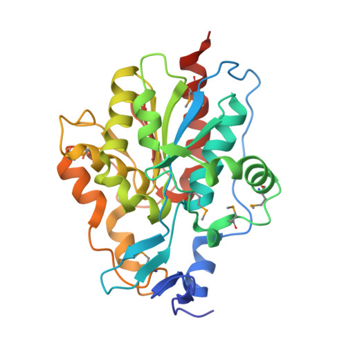
    Heat shock proteins (Hsps) play essential protective roles under stress conditions by preventing the formation of protein aggregates and degrading misfolded proteins. EcHsp31, the yedU (hchA) gene product, is a representative member of a family of chaperones that alleviates protein misfolding by interacting with early unfolding intermediates. The 1.6-A crystal structure of the EcHsp31 dimer reveals a system of hydrophobic patches, canyons, and grooves, which may stabilize partially unfolded substrate. The presence of a well conserved, yet buried, triad in each two-domain subunit suggests a still unproven hydrolytic function of the protein. A flexible extended linker between the A and P domains may play a role in conformational flexibility and substrate binding. The alpha-beta sandwich of the EcHsp31 monomer shows structural similarity to PhPI, a protease belonging to the DJ-1 superfamily. The structure-guided sequence alignment indicates that Hsp31 homologs can be divided in three classes based on variations in the P domain that dramatically affect both oligomerization and catalytic triad formation.


  • Organizational Affiliation

    Biomolecular Structure Center, P.O. Box 357742, University of Washington, Seattle, WA 98195, USA. paulene@u.washington.edu


Macromolecules
Find similar proteins by:  (by identity cutoff)  |  3D Structure
Entity ID: 1
MoleculeChains Sequence LengthOrganismDetailsImage
Chaperone Hsp31291Escherichia coliMutation(s): 8 
Gene Names: hchA/yedU
UniProt
Find proteins for P31658 (Escherichia coli (strain K12))
Explore P31658 
Go to UniProtKB:  P31658
Entity Groups  
Sequence Clusters30% Identity50% Identity70% Identity90% Identity95% Identity100% Identity
UniProt GroupP31658
Sequence Annotations
Expand
  • Reference Sequence
Small Molecules
Ligands 1 Unique
IDChains Name / Formula / InChI Key2D Diagram3D Interactions
MG
Query on MG

Download Ideal Coordinates CCD File 
B [auth A]MAGNESIUM ION
Mg
JLVVSXFLKOJNIY-UHFFFAOYSA-N
Modified Residues  1 Unique
IDChains TypeFormula2D DiagramParent
MSE
Query on MSE
A
L-PEPTIDE LINKINGC5 H11 N O2 SeMET
Experimental Data & Validation

Experimental Data

  • Method: X-RAY DIFFRACTION
  • Resolution: 1.60 Å
  • R-Value Free: 0.242 
  • R-Value Work: 0.187 
  • R-Value Observed: 0.190 
  • Space Group: C 1 2 1
Unit Cell:
Length ( Å )Angle ( ˚ )
a = 52.15α = 90
b = 82β = 100
c = 64.48γ = 90
Software Package:
Software NamePurpose
HKL-2000data collection
SCALEPACKdata scaling
SOLVEphasing
RESOLVEmodel building
REFMACrefinement
HKL-2000data reduction
RESOLVEphasing

Structure Validation

View Full Validation Report


Entry History 

Deposition Data

Revision History  (Full details and data files)

  • Version 1.0: 2003-03-18
    Type: Initial release
  • Version 1.1: 2008-04-28
    Changes: Version format compliance
  • Version 1.2: 2011-07-13
    Changes: Version format compliance