1MXT

Atomic resolution structure of Cholesterol oxidase (Streptomyces sp. SA-COO)


Experimental Data Snapshot

  • Method: X-RAY DIFFRACTION
  • Resolution: 0.95 Å
  • R-Value Free: 0.132 
  • R-Value Work: 0.110 
  • R-Value Observed: 0.098 

wwPDB Validation   3D Report Full Report


This is version 1.3 of the entry. See complete history


Literature

Sub-atomic resolution crystal structure of cholesterol oxidase: What atomic resolution crystallography reveals about enzyme mechanism and the role of FAD cofactor in redox activity

Lario, P.I.Sampson, N.Vrielink, A.

(2003) J Mol Biol 326: 1635-1650

  • DOI: https://doi.org/10.1016/s0022-2836(03)00054-8
  • Primary Citation of Related Structures:  
    1MXT

  • PubMed Abstract: 

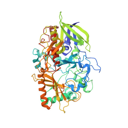
    The crystal structure of cholesterol oxidase, a 56kDa flavoenzyme was anisotropically refined to 0.95A resolution. The final crystallographic R-factor and R(free) value is 11.0% and 13.2%, respectively. The quality of the electron density maps has enabled modeling of alternate conformations for 83 residues in the enzyme, many of which are located in the active site. The additional observed structural features were not apparent in the previous high-resolution structure (1.5A resolution) and have enabled the identification of a narrow tunnel leading directly to the isoalloxazine portion of the FAD prosthetic group. The hydrophobic nature of this narrow tunnel suggests it is the pathway for molecular oxygen to access the isoalloxazine group for the oxidative half reaction. Resolving the alternate conformations in the active site residues provides a model for the dynamics of substrate binding and a potential oxidation triggered gating mechanism involving access to the hydrophobic tunnel. This structure reveals that the NE2 atom of the active site histidine residue, H447, critical to the redox activity of this flavin oxidase, acts as a hydrogen bond donor rather than as hydrogen acceptor. The atomic resolution structure of cholesterol oxidase has revealed the presence of hydrogen atoms, dynamic aspects of the protein and how side-chain conformations are correlated with novel structural features such as the oxygen tunnel. This new structural information has provided us with the opportunity to re-analyze the roles played by specific residues in the mechanism of the enzyme.


  • Organizational Affiliation

    Department of Molecular, Cellular and Developmental Biology, Sinsheimer Laboratory, University of California Santa Cruz, 1156 High Street, Santa Cruz, CA 95064, USA.


Macromolecules
Find similar proteins by:  (by identity cutoff)  |  3D Structure
Entity ID: 1
MoleculeChains Sequence LengthOrganismDetailsImage
CHOLESTEROL OXIDASE504Streptomyces sp.Mutation(s): 0 
Gene Names: CHOA
EC: 1.1.3.6
UniProt
Find proteins for P12676 (Streptomyces sp. (strain SA-COO))
Explore P12676 
Go to UniProtKB:  P12676
Entity Groups  
Sequence Clusters30% Identity50% Identity70% Identity90% Identity95% Identity100% Identity
UniProt GroupP12676
Sequence Annotations
Expand
  • Reference Sequence
Small Molecules
Ligands 3 Unique
IDChains Name / Formula / InChI Key2D Diagram3D Interactions
FAE
Query on FAE

Download Ideal Coordinates CCD File 
C [auth A]FLAVIN-N7 PROTONATED-ADENINE DINUCLEOTIDE
C27 H34 N9 O15 P2
VWWQXMAJTJZDQX-UYBVJOGSSA-O
SO4
Query on SO4

Download Ideal Coordinates CCD File 
B [auth A]SULFATE ION
O4 S
QAOWNCQODCNURD-UHFFFAOYSA-L
OXY
Query on OXY

Download Ideal Coordinates CCD File 
D [auth A]OXYGEN MOLECULE
O2
MYMOFIZGZYHOMD-UHFFFAOYSA-N
Experimental Data & Validation

Experimental Data

  • Method: X-RAY DIFFRACTION
  • Resolution: 0.95 Å
  • R-Value Free: 0.132 
  • R-Value Work: 0.110 
  • R-Value Observed: 0.098 
  • Space Group: P 1 21 1
Unit Cell:
Length ( Å )Angle ( ˚ )
a = 51.227α = 90
b = 72.902β = 105.1
c = 62.947γ = 90
Software Package:
Software NamePurpose
DENZOdata reduction
SCALEPACKdata scaling
SHELXmodel building
SHELXL-97refinement
SHELXphasing

Structure Validation

View Full Validation Report


Entry History 

Deposition Data

Revision History  (Full details and data files)

  • Version 1.0: 2003-02-25
    Type: Initial release
  • Version 1.1: 2008-04-28
    Changes: Version format compliance
  • Version 1.2: 2011-07-13
    Changes: Version format compliance
  • Version 1.3: 2023-09-13
    Changes: Advisory, Data collection, Database references, Derived calculations, Refinement description