Dissociative phosphoryl transfer in PEP mutase catalysis: structure of the enzyme/sulfopyruvate complex and kinetic properties of mutants.
Liu, S., Lu, Z., Jia, Y., Dunaway-Mariano, D., Herzberg, O.(2002) Biochemistry 41: 10270-10276
- PubMed: 12162742
- DOI: https://doi.org/10.1021/bi026024v
- Primary Citation of Related Structures:
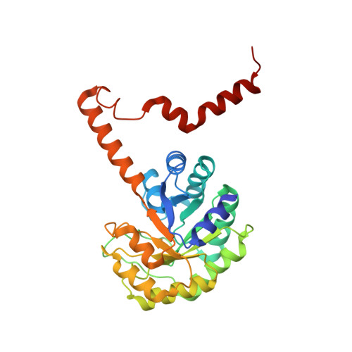
1M1B - PubMed Abstract:
The crystal structure of PEP mutase from Mytilus edulis in complex with a substrate-analogue inhibitor, sulfopyruvate S-pyr (K(i) = 22 microM), has been determined at 2.25 A resolution. Mg(II)-S-pyr binds in the alpha/beta barrel's central channel, at the C-termini of the beta-strands. The binding mode of S-pyr's pyruvyl moiety resembles the binding mode of oxalate seen earlier. The location of the sulfo group of S-pyr is postulated to mimic the phosphonyl group of the product phosphonopyruvate (P-pyr). This sulfo group interacts with the guanidinium group of Arg159, but it is not aligned for nucleopilic attack by neighboring basic amino side chains. Kinetic analysis of site directed mutants, probing the key active site residues Asp58, Arg159, Asn122, and His190 correlate well with the structural information. The results presented here rule out a phosphoryl transfer mechanism involving a double displacement, and suggest instead that PEP mutase catalysis proceeds via a dissociative mechanism in which the pyruvyl C(3) adds to the same face of the phosphorus from which the C(2)O departs. We propose that Arg159 and His190 serve to hold the phosphoryl/metaphosphate/phosphonyl group stationary along the reaction pathway, while the pyruvyl C(1)-C(2) bond rotates upon formation of the metaphosphate. In agreement with published data, the phosphoryl group transfer occurs on the Si-face of PEP with retention of configuration at phosphorus.
Organizational Affiliation:
Center for Advanced Research in Biotechnology, University of Maryland Biotechnology Institute, 9600 Gudelsky Drive, Rockville, Maryland 20850, USA.