Structural characterization of the closed conformation of mouse guanylate kinase.
Sekulic, N., Shuvalova, L., Spangenberg, O., Konrad, M., Lavie, A.(2002) J Biol Chem 277: 30236-30243
- PubMed: 12036965
- DOI: https://doi.org/10.1074/jbc.M204668200
- Primary Citation of Related Structures:
1LVG - PubMed Abstract:
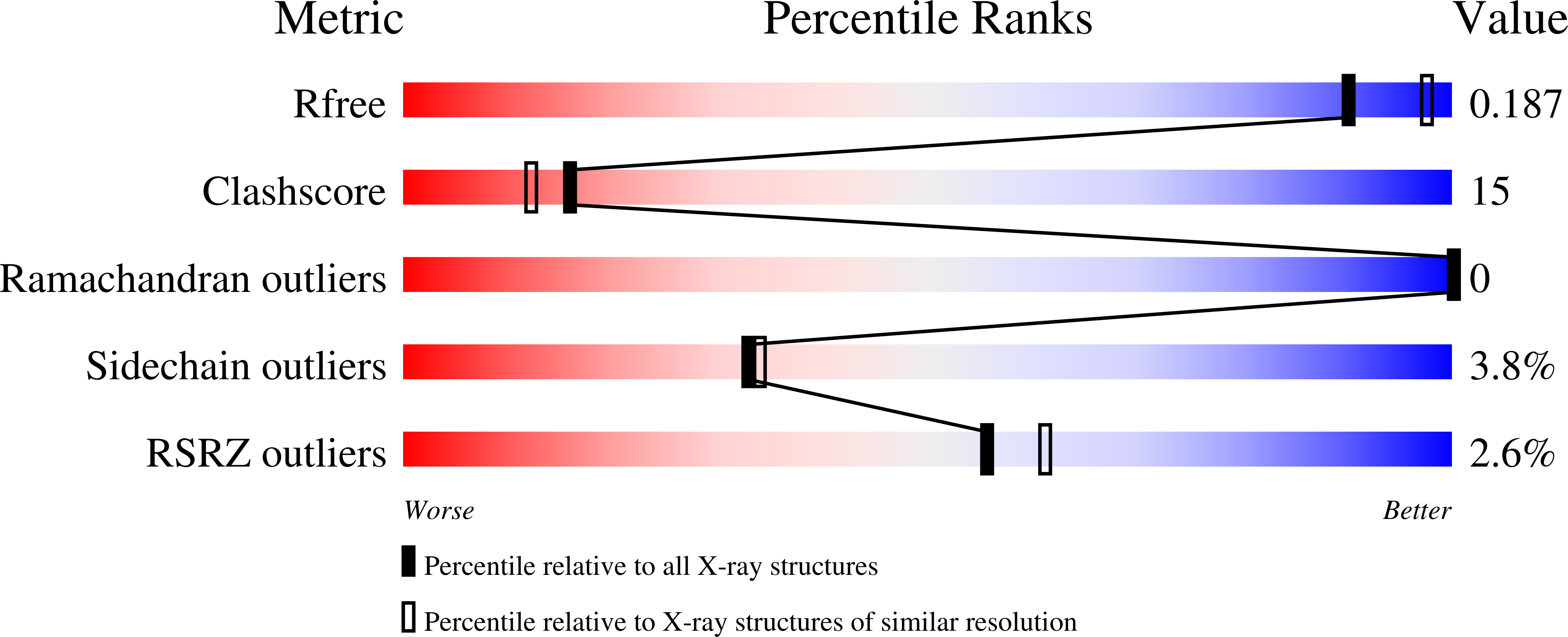
Guanylate kinase (GMPK) is a nucleoside monophosphate kinase that catalyzes the reversible phosphoryl transfer from ATP to GMP to yield ADP and GDP. In addition to phosphorylating GMP, antiviral prodrugs such as acyclovir, ganciclovir, and carbovir and anticancer prodrugs such as the thiopurines are dependent on GMPK for their activation. Hence, structural information on mammalian GMPK could play a role in the design of improved antiviral and antineoplastic agents. Here we present the structure of the mouse enzyme in an abortive complex with the nucleotides ADP and GMP, refined at 2.1 A resolution with a final crystallographic R factor of 0.19 (R(free) = 0.23). Guanylate kinase is a member of the nucleoside monophosphate (NMP) kinase family, a family of enzymes that despite having a low primary structure identity share a similar fold, which consists of three structurally distinct regions termed the CORE, LID, and NMP-binding regions. Previous studies on the yeast enzyme have shown that these parts move as rigid bodies upon substrate binding. It has been proposed that consecutive binding of substrates leads to "closing" of the active site bringing the NMP-binding and LID regions closer to each other and to the CORE region. Our structure, which is the first of any guanylate kinase with both substrates bound, supports this hypothesis. It also reveals the binding site of ATP and implicates arginines 44, 137, and 148 (in addition to the invariant P-loop lysine) as candidates for catalyzing the chemical step of the phosphoryl transfer.
Organizational Affiliation:
University of Illinois at Chicago, Department of Biochemistry and Molecular Biology, Chicago, Illinois 60612 and the Max Planck Institute for Biophysical Chemistry, Department of Molecular Genetics, Am Fassberg 11, 37077 Göttingen, Germany.


