1J2E

Crystal structure of Human Dipeptidyl peptidase IV


Experimental Data Snapshot

  • Method: X-RAY DIFFRACTION
  • Resolution: 2.60 Å
  • R-Value Free: 0.302 
  • R-Value Work: 0.249 

wwPDB Validation   3D Report Full Report


This is version 1.4 of the entry. See complete history


Literature

The structure and function of human dipeptidyl peptidase IV, possessing a unique eight-bladed beta-propeller fold.

Hiramatsu, H.Kyono, K.Higashiyama, Y.Fukushima, C.Shima, H.Sugiyama, S.Inaka, K.Yamamoto, A.Shimizu, R.

(2003) Biochem Biophys Res Commun 302: 849-854

  • DOI: https://doi.org/10.1016/s0006-291x(03)00258-4
  • Primary Citation of Related Structures:  
    1J2E

  • PubMed Abstract: 

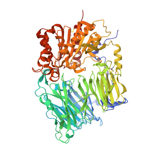
    Dipeptidyl peptidase IV (DPPIV) is a serine protease, a member of the prolyl oligopeptidase (POP) family, and has been implicated in several diseases. Therefore, the development of DPPIV selective inhibitors, which are able to control the biological function of DPPIV, is important. We determined the crystal structure of human DPPIV at 2.6A resolution. The molecule consists of a unique eight-bladed beta-propeller domain in the N-terminal region and a serine protease domain in the C-terminal region. Also, the large "cave" structure, which is thought to control the access of the substrate, is found on the side of the beta-propeller fold. Comparison of the overall amino acid sequence between human DPPIV and POP shows low homology (12.9%). In this paper, we report the structure of human DPPIV, especially focusing on a unique eight-bladed beta-propeller domain. We also discuss the way for the access of the substrate to this domain.


  • Organizational Affiliation

    Discovery Research Laboratory, Tanabe Seiyaku Co. Ltd., 3-16-86 Kashima, Yodogawa-ku, Osaka 532-8505, Japan.


Macromolecules
Find similar proteins by:  (by identity cutoff)  |  3D Structure
Entity ID: 1
MoleculeChains Sequence LengthOrganismDetailsImage
Dipeptidyl peptidase IV
A, B
740Homo sapiensMutation(s): 0 
EC: 3.4.14.5
UniProt & NIH Common Fund Data Resources
Find proteins for P27487 (Homo sapiens)
Explore P27487 
Go to UniProtKB:  P27487
PHAROS:  P27487
GTEx:  ENSG00000197635 
Entity Groups  
Sequence Clusters30% Identity50% Identity70% Identity90% Identity95% Identity100% Identity
UniProt GroupP27487
Sequence Annotations
Expand
  • Reference Sequence
Experimental Data & Validation

Experimental Data

  • Method: X-RAY DIFFRACTION
  • Resolution: 2.60 Å
  • R-Value Free: 0.302 
  • R-Value Work: 0.249 
  • Space Group: P 21 21 21
Unit Cell:
Length ( Å )Angle ( ˚ )
a = 118.04α = 90
b = 125.92β = 90
c = 136.84γ = 90
Software Package:
Software NamePurpose
MOSFLMdata reduction
SCALAdata scaling
MLPHAREphasing
CNXrefinement
CCP4data scaling

Structure Validation

View Full Validation Report


Entry History 

Deposition Data

Revision History  (Full details and data files)

  • Version 1.0: 2003-12-30
    Type: Initial release
  • Version 1.1: 2008-04-27
    Changes: Version format compliance
  • Version 1.2: 2011-07-13
    Changes: Non-polymer description, Version format compliance
  • Version 1.3: 2020-07-29
    Type: Remediation
    Reason: Carbohydrate remediation
    Changes: Data collection, Database references, Derived calculations, Structure summary
  • Version 1.4: 2023-12-27
    Changes: Data collection, Database references, Structure summary