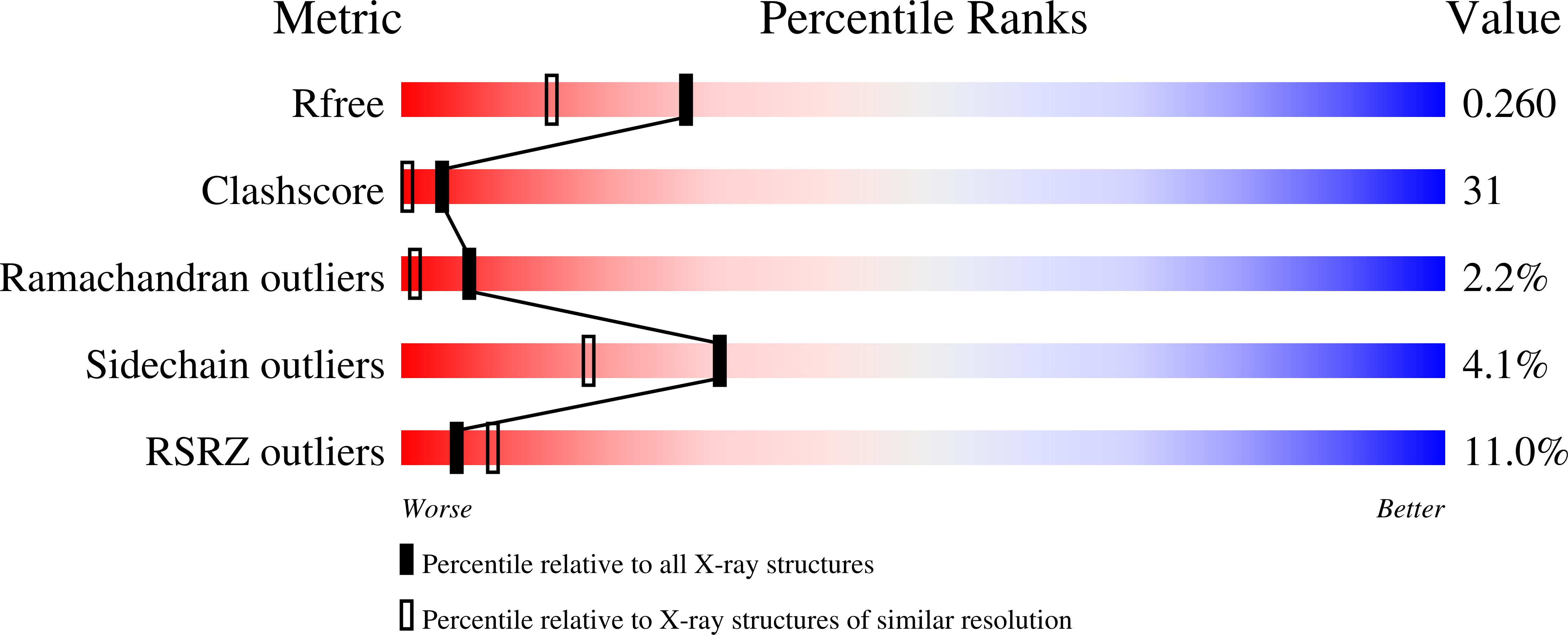
1I7E

C-Terminal Domain Of Mouse Brain Tubby Protein bound to Phosphatidylinositol 4,5-bis-phosphate


Experimental Data Snapshot

  • Method: X-RAY DIFFRACTION
  • Resolution: 1.95 Å
  • R-Value Free: 0.261 
  • R-Value Work: 0.212 
  • R-Value Observed: 0.212 

wwPDB Validation   3D Report Full Report


Ligand Structure Quality Assessment 


This is version 1.4 of the entry. See complete history


Literature

G-protein signaling through tubby proteins.

Santagata, S.Boggon, T.J.Baird, C.L.Gomez, C.A.Zhao, J.Shan, W.S.Myszka, D.G.Shapiro, L.

(2001) Science 292: 2041-2050

  • DOI: https://doi.org/10.1126/science.1061233
  • Primary Citation of Related Structures:  
    1I7E

  • PubMed Abstract: 

    Dysfunction of the tubby protein results in maturity-onset obesity in mice. Tubby has been implicated as a transcription regulator, but details of the molecular mechanism underlying its function remain unclear. Here we show that tubby functions in signal transduction from heterotrimeric GTP-binding protein (G protein)-coupled receptors. Tubby localizes to the plasma membrane by binding phosphatidylinositol 4,5-bisphosphate through its carboxyl terminal "tubby domain." X-ray crystallography reveals the atomic-level basis of this interaction and implicates tubby domains as phosphorylated-phosphatidyl- inositol binding factors. Receptor-mediated activation of G protein alphaq (Galphaq) releases tubby from the plasma membrane through the action of phospholipase C-beta, triggering translocation of tubby to the cell nucleus. The localization of tubby-like protein 3 (TULP3) is similarly regulated. These data suggest that tubby proteins function as membrane-bound transcription regulators that translocate to the nucleus in response to phosphoinositide hydrolysis, providing a direct link between G-protein signaling and the regulation of gene expression.


  • Organizational Affiliation

    Ruttenberg Cancer Center, Structural Biology Program, Department of Physiology and Biophysics, Mount Sinai School of Medicine of New York University, 1425 Madison Avenue New York, NY 10029, USA.


Macromolecules
Find similar proteins by:  (by identity cutoff)  |  3D Structure
Entity ID: 1
MoleculeChains Sequence LengthOrganismDetailsImage
TUBBY PROTEIN265Mus musculusMutation(s): 0 
Membrane Entity: Yes 
UniProt
Find proteins for P50586 (Mus musculus)
Explore P50586 
Go to UniProtKB:  P50586
Entity Groups  
Sequence Clusters30% Identity50% Identity70% Identity90% Identity95% Identity100% Identity
UniProt GroupP50586
Sequence Annotations
Expand
  • Reference Sequence
Small Molecules
Ligands 1 Unique
IDChains Name / Formula / InChI Key2D Diagram3D Interactions
IBS
Query on IBS

Download Ideal Coordinates CCD File 
B [auth A]L-ALPHA-GLYCEROPHOSPHO-D-MYO-INOSITOL-4,5-BIS-PHOSPHATE
C9 H19 O17 P3
JBQPYAMQMBKZDT-WKBARDANSA-N
Experimental Data & Validation

Experimental Data

  • Method: X-RAY DIFFRACTION
  • Resolution: 1.95 Å
  • R-Value Free: 0.261 
  • R-Value Work: 0.212 
  • R-Value Observed: 0.212 
  • Space Group: P 21 21 21
Unit Cell:
Length ( Å )Angle ( ˚ )
a = 43.102α = 90
b = 51.469β = 90
c = 120.834γ = 90
Software Package:
Software NamePurpose
X-PLORmodel building
CNSrefinement
DENZOdata reduction
SCALEPACKdata scaling
X-PLORphasing

Structure Validation

View Full Validation Report


Ligand Structure Quality Assessment 


Entry History 

Deposition Data

Revision History  (Full details and data files)

  • Version 1.0: 2001-06-27
    Type: Initial release
  • Version 1.1: 2008-04-27
    Changes: Version format compliance
  • Version 1.2: 2011-07-13
    Changes: Version format compliance
  • Version 1.3: 2018-01-31
    Changes: Experimental preparation
  • Version 1.4: 2023-08-09
    Changes: Data collection, Database references, Derived calculations, Refinement description