1FX8

CRYSTAL STRUCTURE OF THE E. COLI GLYCEROL FACILITATOR (GLPF) WITH SUBSTRATE GLYCEROL


Experimental Data Snapshot

  • Method: X-RAY DIFFRACTION
  • Resolution: 2.20 Å
  • R-Value Free: 0.223 
  • R-Value Work: 0.197 
  • R-Value Observed: 0.197 

wwPDB Validation   3D Report Full Report


This is version 1.4 of the entry. See complete history


Literature

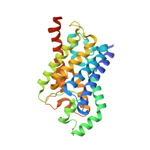
Structure of a glycerol-conducting channel and the basis for its selectivity.

Fu, D.Libson, A.Miercke, L.J.Weitzman, C.Nollert, P.Krucinski, J.Stroud, R.M.

(2000) Science 290: 481-486

  • DOI: https://doi.org/10.1126/science.290.5491.481
  • Primary Citation of Related Structures:  
    1FX8

  • PubMed Abstract: 

    Membrane channel proteins of the aquaporin family are highly selective for permeation of specific small molecules, with absolute exclusion of ions and charged solutes and without dissipation of the electrochemical potential across the cell membrane. We report the crystal structure of the Escherichia coli glycerol facilitator (GlpF) with its primary permeant substrate glycerol at 2.2 angstrom resolution. Glycerol molecules line up in an amphipathic channel in single file. In the narrow selectivity filter of the channel the glycerol alkyl backbone is wedged against a hydrophobic corner, and successive hydroxyl groups form hydrogen bonds with a pair of acceptor, and donor atoms. Two conserved aspartic acid-proline-alanine motifs form a key interface between two gene-duplicated segments that each encode three-and-one-half membrane-spanning helices around the channel. This structure elucidates the mechanism of selective permeability for linear carbohydrates and suggests how ions and water are excluded.


  • Organizational Affiliation

    Department of Biochemistry and Biophysics, School of Medicine, University of California, San Francisco, CA 94143-0448, USA.


Macromolecules
Find similar proteins by:  (by identity cutoff)  |  3D Structure
Entity ID: 1
MoleculeChains Sequence LengthOrganismDetailsImage
GLYCEROL UPTAKE FACILITATOR PROTEIN281Escherichia coliMutation(s): 0 
Membrane Entity: Yes 
UniProt
Find proteins for P0AER0 (Escherichia coli (strain K12))
Explore P0AER0 
Go to UniProtKB:  P0AER0
Entity Groups  
Sequence Clusters30% Identity50% Identity70% Identity90% Identity95% Identity100% Identity
UniProt GroupP0AER0
Sequence Annotations
Expand
  • Reference Sequence
Experimental Data & Validation

Experimental Data

  • Method: X-RAY DIFFRACTION
  • Resolution: 2.20 Å
  • R-Value Free: 0.223 
  • R-Value Work: 0.197 
  • R-Value Observed: 0.197 
  • Space Group: I 4 2 2
Unit Cell:
Length ( Å )Angle ( ˚ )
a = 96.76α = 90
b = 96.76β = 90
c = 184.322γ = 90
Software Package:
Software NamePurpose
SHARPphasing
CNSrefinement
MOSFLMdata reduction
CCP4data scaling

Structure Validation

View Full Validation Report


Entry History 

Deposition Data

Revision History  (Full details and data files)

  • Version 1.0: 2000-11-01
    Type: Initial release
  • Version 1.1: 2008-04-27
    Changes: Version format compliance
  • Version 1.2: 2011-07-13
    Changes: Derived calculations, Version format compliance
  • Version 1.3: 2020-07-29
    Type: Remediation
    Reason: Carbohydrate remediation
    Changes: Data collection, Derived calculations, Structure summary
  • Version 1.4: 2024-02-07
    Changes: Data collection, Database references, Structure summary