1BI4

CATALYTIC DOMAIN OF HIV-1 INTEGRASE


Experimental Data Snapshot

  • Method: X-RAY DIFFRACTION
  • Resolution: 2.50 Å
  • R-Value Free: 0.271 
  • R-Value Work: 0.232 
  • R-Value Observed: 0.232 

wwPDB Validation   3D Report Full Report


This is version 1.4 of the entry. See complete history


Literature

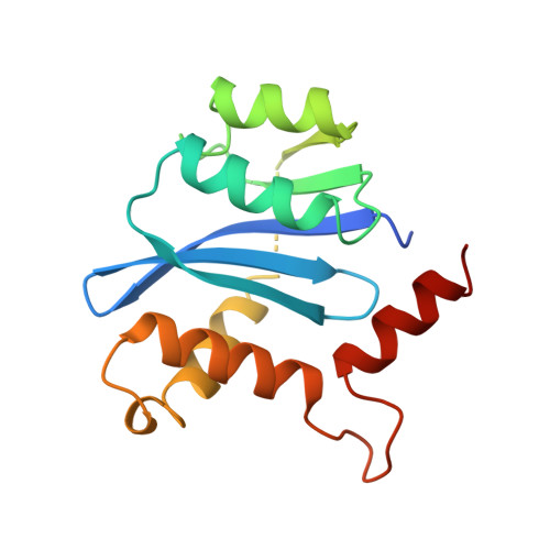
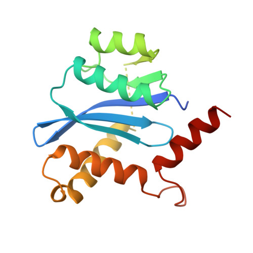
Crystal structures of the catalytic domain of HIV-1 integrase free and complexed with its metal cofactor: high level of similarity of the active site with other viral integrases.

Maignan, S.Guilloteau, J.P.Zhou-Liu, Q.Clement-Mella, C.Mikol, V.

(1998) J Mol Biol 282: 359-368

  • DOI: https://doi.org/10.1006/jmbi.1998.2002
  • Primary Citation of Related Structures:  
    1BHL, 1BI4, 1BL3

  • PubMed Abstract: 

    Human immunodeficiency virus (HIV) integrase is the enzyme responsible for insertion of a DNA copy of the viral genome into host DNA, an essential step in the replication cycle of HIV. HIV-1 integrase comprises three functional and structural domains: an N-terminal zinc-binding domain, a catalytic core domain and a C-terminal DNA-binding domain. The catalytic core domain with the F185H mutation has been crystallized without sodium cacodylate in a new crystal form, free and complexed with the catalytic metal Mg2+. The structures have been determined and refined to about 2.2 A. Unlike the previously reported structures, the three active-site carboxylate residues (D,D-35-E motif) are well ordered and both aspartate residues delineate a proper metal-binding site. Comparison of the active binding site of this domain with that of other members from the polynucleotidyl transferases superfamily shows a high level of similarity, providing a confident template for the design of antiviral agents.


  • Organizational Affiliation

    Rhône-Poulenc Rorer, 13, Quai J. Guesde, Vitry/Seine, F-94403, France.


Macromolecules
Find similar proteins by:  (by identity cutoff)  |  3D Structure
Entity ID: 1
MoleculeChains Sequence LengthOrganismDetailsImage
INTEGRASE
A, C
160Human immunodeficiency virus 1Mutation(s): 1 
UniProt
Find proteins for P12497 (Human immunodeficiency virus type 1 group M subtype B (isolate NY5))
Explore P12497 
Go to UniProtKB:  P12497
Entity Groups  
Sequence Clusters30% Identity50% Identity70% Identity90% Identity95% Identity100% Identity
UniProt GroupP12497
Sequence Annotations
Expand
  • Reference Sequence
Find similar proteins by:  (by identity cutoff)  |  3D Structure
Entity ID: 2
MoleculeChains Sequence LengthOrganismDetailsImage
INTEGRASE160Human immunodeficiency virus 1Mutation(s): 1 
UniProt
Find proteins for P12497 (Human immunodeficiency virus type 1 group M subtype B (isolate NY5))
Explore P12497 
Go to UniProtKB:  P12497
Entity Groups  
Sequence Clusters30% Identity50% Identity70% Identity90% Identity95% Identity100% Identity
UniProt GroupP12497
Sequence Annotations
Expand
  • Reference Sequence
Experimental Data & Validation

Experimental Data

  • Method: X-RAY DIFFRACTION
  • Resolution: 2.50 Å
  • R-Value Free: 0.271 
  • R-Value Work: 0.232 
  • R-Value Observed: 0.232 
  • Space Group: C 1 2 1
Unit Cell:
Length ( Å )Angle ( ˚ )
a = 99.1α = 90
b = 72.5β = 108
c = 79.4γ = 90
Software Package:
Software NamePurpose
X-PLORmodel building
X-PLORrefinement
DENZOdata reduction
SCALEPACKdata scaling
X-PLORphasing

Structure Validation

View Full Validation Report


Entry History 

Deposition Data

Revision History  (Full details and data files)

  • Version 1.0: 1998-11-04
    Type: Initial release
  • Version 1.1: 2008-03-24
    Changes: Version format compliance
  • Version 1.2: 2011-07-13
    Changes: Version format compliance
  • Version 1.3: 2021-11-03
    Changes: Database references, Other
  • Version 1.4: 2023-08-02
    Changes: Refinement description