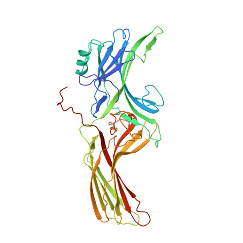
1AYR

ARRESTIN FROM BOVINE ROD OUTER SEGMENTS


Experimental Data Snapshot

  • Method: X-RAY DIFFRACTION
  • Resolution: 3.30 Å
  • R-Value Free: 0.316 
  • R-Value Work: 0.254 
  • R-Value Observed: 0.254 

wwPDB Validation   3D Report Full Report


This is version 1.3 of the entry. See complete history


Literature

X-ray crystal structure of arrestin from bovine rod outer segments.

Granzin, J.Wilden, U.Choe, H.W.Labahn, J.Krafft, B.Buldt, G.

(1998) Nature 391: 918-921

  • DOI: https://doi.org/10.1038/36147
  • Primary Citation of Related Structures:  
    1AYR

  • PubMed Abstract: 

    Retinal arrestin is the essential protein for the termination of the light response in vertebrate rod outer segments. It plays an important role in quenching the light-induced enzyme cascade by its ability to bind to phosphorylated light-activated rhodopsin (P-Rh*). Arrestins are found in various G-protein-coupled amplification cascades. Here we report on the three-dimensional structure of bovine arrestin (relative molecular mass, 45,300) at 3.3 A resolution. The crystal structure comprises two domains of antiparallel beta-sheets connected through a hinge region and one short alpha-helix on the back of the amino-terminal fold. The binding region for phosphorylated light-activated rhodopsin is located at the N-terminal domain, as indicated by the docking of the photoreceptor to the three-dimensional structure of arrestin. This agrees with the interpretation of binding studies on partially digested and mutated arrestin.


  • Organizational Affiliation

    Forschungszentrum Jülich, Institut für Biologische Informationsverarbeitung, Germany. J.Granzin@fz-juelich.de


Macromolecules
Find similar proteins by:  (by identity cutoff)  |  3D Structure
Entity ID: 1
MoleculeChains Sequence LengthOrganismDetailsImage
ARRESTIN
A, B, C, D
368Bos taurusMutation(s): 0 
UniProt
Find proteins for P08168 (Bos taurus)
Explore P08168 
Go to UniProtKB:  P08168
Entity Groups  
Sequence Clusters30% Identity50% Identity70% Identity90% Identity95% Identity100% Identity
UniProt GroupP08168
Sequence Annotations
Expand
  • Reference Sequence
Experimental Data & Validation

Experimental Data

  • Method: X-RAY DIFFRACTION
  • Resolution: 3.30 Å
  • R-Value Free: 0.316 
  • R-Value Work: 0.254 
  • R-Value Observed: 0.254 
  • Space Group: P 21 21 2
Unit Cell:
Length ( Å )Angle ( ˚ )
a = 169.17α = 90
b = 185.53β = 90
c = 90.93γ = 90
Software Package:
Software NamePurpose
X-PLORmodel building
X-PLORrefinement
DENZOdata reduction
CCP4data scaling
X-PLORphasing

Structure Validation

View Full Validation Report


Entry History 

Deposition Data

Revision History  (Full details and data files)

  • Version 1.0: 1998-11-25
    Type: Initial release
  • Version 1.1: 2008-03-03
    Changes: Version format compliance
  • Version 1.2: 2011-07-13
    Changes: Version format compliance
  • Version 1.3: 2024-02-07
    Changes: Data collection, Database references, Other