High-resolution Cryo-EM Structure Determination of a-Synuclein-A Prototypical Amyloid Fibril.
Sanchez, J.C., Pierson, J.A., Borcik, C.G., Rienstra, C.M., Wright, E.R.(2025) Bio Protoc 15: e5171-e5171
- PubMed: 39959285 Search on PubMedSearch on PubMed Central
- DOI: https://doi.org/10.21769/BioProtoc.5171
- Primary Citation Related Structures:
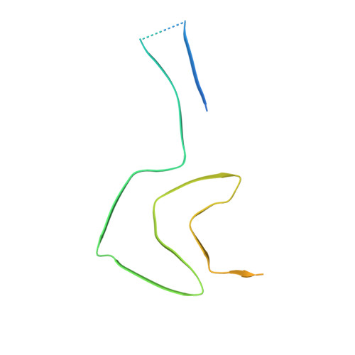
9CK3 - PubMed Abstract:
The physiological role of a-synuclein (a-syn), an intrinsically disordered presynaptic neuronal protein, is believed to impact the release of neurotransmitters through interactions with the SNARE complex. However, under certain cellular conditions that are not well understood, a-syn will self-assemble into β-sheet-rich fibrils that accumulate and form insoluble neuronal inclusions. Studies of patient-derived brain tissues have concluded that these inclusions are associated with Parkinson's disease, the second most common neurodegenerative disorder, and other synuclein-related diseases called synucleinopathies. In addition, repetitions of specific mutations to the SNCA gene, the gene that encodes a-syn, result in an increased disposition for synucleinopathies. The latest advances in cryo-EM structure determination and real-space helical reconstruction methods have resulted in over 60 in vitro structures of a-syn fibrils solved to date, with a handful of these reaching a resolution below 2.5 Å. Here, we provide a protocol for a-syn protein expression, purification, and fibrilization. We detail how sample quality is assessed by negative stain transmission electron microscopy (NS-TEM) analysis and followed by sample vitrification using the Vitrobot Mark IV vitrification robot. We provide a detailed step-by-step protocol for high-resolution cryo-EM structure determination of a-syn fibrils using RELION and a series of specialized helical reconstruction tools that can be run within RELION. Finally, we detail how ChimeraX, Coot, and Phenix are used to build and refine a molecular model into the high-resolution cryo-EM map. This workflow resulted in a 2.04 Å structure of a-syn fibrils with excellent resolution of residues 36-97 and an additional island of density for residues 15-22 that had not been previously reported. This workflow should serve as a starting point for individuals new to the neurodegeneration and structural biology fields. Together, this procedure lays the foundation for advanced structural studies of a-syn and other amyloid fibrils. Key features • In vitro fibril amplification method yielding twisting fibrils that span several micrometers in length and are suitable for cryo-EM structure determination. • High-throughput cryo-EM data collection of neurodegenerative fibrils, such as alpha-synuclein. • Use of RELION implementations of helical reconstruction algorithms to generate high-resolution 3D structures of a-synuclein fibrils. • Brief demonstration of the use of ChimeraX, Coot, and Phenix for molecular model building and refinement.
- Department of Biochemistry, University of Wisconsin-Madison, Madison, WI, USA.
Organizational Affiliation: