6YLO

mTurquoise2 - Directionality of Optical Properties of Fluorescent Proteins


Experimental Data Snapshot

  • Method: X-RAY DIFFRACTION
  • Resolution: 1.70 Å
  • R-Value Free: 0.205 
  • R-Value Work: 0.179 
  • R-Value Observed: 0.180 

wwPDB Validation   3D Report Full Report


This is version 1.2 of the entry. See complete history


Literature

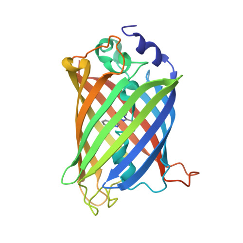
Directionality of light absorption and emission in representative fluorescent proteins.

Myskova, J.Rybakova, O.Brynda, J.Khoroshyy, P.Bondar, A.Lazar, J.

(2020) Proc Natl Acad Sci U S A 117: 32395-32401

  • DOI: https://doi.org/10.1073/pnas.2017379117
  • Primary Citation of Related Structures:  
    6YLM, 6YLN, 6YLO, 6YLP, 6YLQ, 6YLS

  • PubMed Abstract: 

    Fluorescent molecules are like antennas: The rate at which they absorb light depends on their orientation with respect to the incoming light wave, and the apparent intensity of their emission depends on their orientation with respect to the observer. However, the directions along which the most important fluorescent molecules in biology, fluorescent proteins (FPs), absorb and emit light are generally not known. Our optical and X-ray investigations of FP crystals have now allowed us to determine the molecular orientations of the excitation and emission transition dipole moments in the FPs mTurquoise2, eGFP, and mCherry, and the photoconvertible FP mEos4b. Our results will allow using FP directionality in studies of molecular and biological processes, but also in development of novel bioengineering and bioelectronics applications.


  • Organizational Affiliation

    Institute of Organic Chemistry and Biochemistry, Czech Academy of Sciences, 16610 Prague 6, Czech Republic.


Macromolecules
Find similar proteins by:  (by identity cutoff)  |  3D Structure
Entity ID: 1
MoleculeChains Sequence LengthOrganismDetailsImage
mTurquoise2_C2221243synthetic constructMutation(s): 0 
Entity Groups  
Sequence Clusters30% Identity50% Identity70% Identity90% Identity95% Identity100% Identity
Sequence Annotations
Expand
  • Reference Sequence
Small Molecules
Modified Residues  1 Unique
IDChains TypeFormula2D DiagramParent
SWG
Query on SWG
A
L-PEPTIDE LINKINGC16 H16 N4 O4SER, TRP, GLY
Experimental Data & Validation

Experimental Data

  • Method: X-RAY DIFFRACTION
  • Resolution: 1.70 Å
  • R-Value Free: 0.205 
  • R-Value Work: 0.179 
  • R-Value Observed: 0.180 
  • Space Group: C 2 2 21
Unit Cell:
Length ( Å )Angle ( ˚ )
a = 60.82α = 90
b = 90.55β = 90
c = 117.85γ = 90
Software Package:
Software NamePurpose
XDSdata reduction
XSCALEdata scaling
MOLREPphasing
REFMACrefinement
PDB_EXTRACTdata extraction

Structure Validation

View Full Validation Report


Entry History & Funding Information

Deposition Data


Funding OrganizationLocationGrant Number
Ministry of Education, Youth and Sports of the Czech RepublicCzech RepublicCZ.02.1.01/0.0/0.0/16_019/0000729

Revision History  (Full details and data files)

  • Version 1.0: 2020-12-16
    Type: Initial release
  • Version 1.1: 2021-01-27
    Changes: Database references
  • Version 1.2: 2024-01-24
    Changes: Data collection, Database references, Derived calculations, Refinement description