6OS6

Crystal structure of CymD prenyltransferase complexed with L-tryptophan and DMSPP


Experimental Data Snapshot

  • Method: X-RAY DIFFRACTION
  • Resolution: 1.33 Å
  • R-Value Free: 0.181 
  • R-Value Work: 0.166 
  • R-Value Observed: 0.167 

wwPDB Validation   3D Report Full Report


Ligand Structure Quality Assessment 


This is version 2.2 of the entry. See complete history


Literature

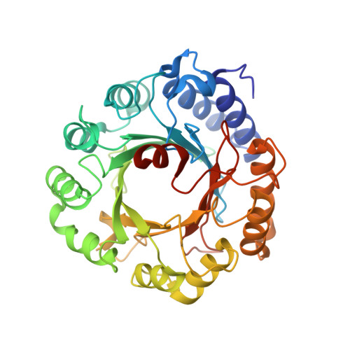
Structural Basis of Tryptophan Reverse N-Prenylation Catalyzed by CymD.

Roose, B.W.Christianson, D.W.

(2019) Biochemistry 58: 3232-3242

  • DOI: https://doi.org/10.1021/acs.biochem.9b00399
  • Primary Citation of Related Structures:  
    6OS3, 6OS5, 6OS6

  • PubMed Abstract: 

    Indole prenyltransferases catalyze the prenylation of l-tryptophan (l-Trp) and other indoles to produce a diverse set of natural products in bacteria, fungi, and plants, many of which possess useful biological properties. Among this family of enzymes, CymD from Salinispora arenicola catalyzes the reverse N1 prenylation of l-Trp, an unusual reaction given the poor nucleophilicity of the indole nitrogen. CymD utilizes dimethylallyl diphosphate (DMAPP) as the prenyl donor, catalyzing the dissociation of the diphosphate leaving group followed by nucleophilic attack of the indole nitrogen at the tertiary carbon of the dimethylallyl cation. To better understand the structural basis of selective indole N-alkylation reactions in biology, we have determined the X-ray crystal structures of CymD, the CymD-l-Trp complex, and the CymD-l-Trp-DMSPP complex (DMSPP is dimethylallyl S -thiolodiphosphate, an unreactive analogue of DMAPP). The orientation of l-Trp with respect to DMSPP reveals how the active site contour of CymD serves as a template to direct the reverse prenylation of the indole nitrogen. Comparison to PriB, a C6 bacterial indole prenyltransferase, offers further insight regarding the structural basis of regioselective indole prenylation. Isothermal titration calorimetry measurements indicate a synergistic relationship between l-Trp and DMSPP binding. Finally, activity assays demonstrate the selectivity of CymD for l-Trp and indole as prenyl acceptors. Collectively, these data establish a foundation for understanding and engineering the regioselectivity of indole prenylation by members of the prenyltransferase protein family.


  • Organizational Affiliation

    Roy and Diana Vagelos Laboratories, Department of Chemistry , University of Pennsylvania , 231 South 34th Street , Philadelphia , Pennsylvania 19104-6323 , United States.


Macromolecules
Find similar proteins by:  (by identity cutoff)  |  3D Structure
Entity ID: 1
MoleculeChains Sequence LengthOrganismDetailsImage
CymD prenyltransferase375Salinispora arenicola CNS-205Mutation(s): 0 
Gene Names: Sare_4565
UniProt
Find proteins for A8M6W6 (Salinispora arenicola (strain CNS-205))
Explore A8M6W6 
Go to UniProtKB:  A8M6W6
Entity Groups  
Sequence Clusters30% Identity50% Identity70% Identity90% Identity95% Identity100% Identity
UniProt GroupA8M6W6
Sequence Annotations
Expand
  • Reference Sequence
Small Molecules
Ligands 5 Unique
IDChains Name / Formula / InChI Key2D Diagram3D Interactions
DST (Subject of Investigation/LOI)
Query on DST

Download Ideal Coordinates CCD File 
B [auth A]DIMETHYLALLYL S-THIOLODIPHOSPHATE
C5 H12 O6 P2 S
ZWFWSISPSBLNGO-UHFFFAOYSA-N
TRP (Subject of Investigation/LOI)
Query on TRP

Download Ideal Coordinates CCD File 
C [auth A]TRYPTOPHAN
C11 H12 N2 O2
QIVBCDIJIAJPQS-VIFPVBQESA-N
BEZ
Query on BEZ

Download Ideal Coordinates CCD File 
G [auth A]BENZOIC ACID
C7 H6 O2
WPYMKLBDIGXBTP-UHFFFAOYSA-N
ZN
Query on ZN

Download Ideal Coordinates CCD File 
D [auth A],
E [auth A]
ZINC ION
Zn
PTFCDOFLOPIGGS-UHFFFAOYSA-N
CL
Query on CL

Download Ideal Coordinates CCD File 
F [auth A]CHLORIDE ION
Cl
VEXZGXHMUGYJMC-UHFFFAOYSA-M
Binding Affinity Annotations 
IDSourceBinding Affinity
TRP Binding MOAD:  6OS6 Kd: 1100 (nM) from 1 assay(s)
DST Binding MOAD:  6OS6 Kd: 1000 (nM) from 1 assay(s)
Experimental Data & Validation

Experimental Data

  • Method: X-RAY DIFFRACTION
  • Resolution: 1.33 Å
  • R-Value Free: 0.181 
  • R-Value Work: 0.166 
  • R-Value Observed: 0.167 
  • Space Group: I 4
Unit Cell:
Length ( Å )Angle ( ˚ )
a = 129.446α = 90
b = 129.446β = 90
c = 49.777γ = 90
Software Package:
Software NamePurpose
XDSdata reduction
Aimlessdata scaling
PHENIXrefinement
PDB_EXTRACTdata extraction
PHASERphasing

Structure Validation

View Full Validation Report


Ligand Structure Quality Assessment 


Entry History & Funding Information

Deposition Data


Funding OrganizationLocationGrant Number
National Institutes of Health/National Institute of General Medical Sciences (NIH/NIGMS)United StatesGM56838

Revision History  (Full details and data files)

  • Version 1.0: 2019-07-17
    Type: Initial release
  • Version 1.1: 2019-08-14
    Changes: Data collection, Database references
  • Version 2.0: 2019-10-02
    Changes: Atomic model, Author supporting evidence, Data collection, Derived calculations, Non-polymer description, Structure summary
  • Version 2.1: 2020-01-01
    Changes: Author supporting evidence
  • Version 2.2: 2023-10-11
    Changes: Data collection, Database references, Derived calculations, Refinement description, Structure summary