2ZFD

The crystal structure of plant specific calcium binding protein AtCBL2 in complex with the regulatory domain of AtCIPK14


Experimental Data Snapshot

  • Method: X-RAY DIFFRACTION
  • Resolution: 1.20 Å
  • R-Value Free: 0.192 
  • R-Value Work: 0.180 
  • R-Value Observed: 0.180 

wwPDB Validation   3D Report Full Report


This is version 1.2 of the entry. See complete history


Literature

The crystal structure of plant-specific calcium-binding protein AtCBL2 in complex with the regulatory domain of AtCIPK14

Akaboshi, M.Hashimoto, H.Ishida, H.Saijo, S.Koizumi, N.Sato, M.Shimizu, T.

(2008) J Mol Biol 377: 246-257

  • DOI: https://doi.org/10.1016/j.jmb.2008.01.006
  • Primary Citation of Related Structures:  
    2ZFD

  • PubMed Abstract: 

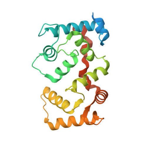
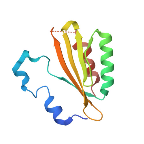
    Calcium signals mediate a multitude of plant responses to external stimuli. Calcineurin B-like (CBL) proteins and their target kinases, CBL-interacting protein kinases (CIPKs), represent important relays in plant calcium signaling. CBL interacts with CIPK through a conserved motif (NAF/FISL motif) within the C-terminal regulatory domain. To better understand the functional role of the CBL-CIPK system, we determined the crystal structure of AtCBL2 in complex with the regulatory domain of AtCIPK14 at 1.2 A resolution. The NAF/FISL motif is inserted into a hydrophobic crevice within AtCBL2, accompanied by a large displacement of the helices and loop on the opposite side of the NAF/FISL motif from the C-terminal region, which shields the hydrophobic crevice in free form. Ca(2+) are coordinated within four EF hands in AtCBL2 in bound form. This calcium coordination pattern differs from that in the structure of the SOS3-SOS2 complex previously reported. Structural comparison of the two structures shows that the recognition of CBL by CIPK is performed in a similar manner, but inherent interactions confer binding affinity and specificity.


  • Organizational Affiliation

    International Graduate School of Arts and Sciences, Yokohama City University, 1-7-29 Suehiro-cho, Tsurumi-ku, Yokohama, Kanagawa 230-0045, Japan.


Macromolecules
Find similar proteins by:  (by identity cutoff)  |  3D Structure
Entity ID: 1
MoleculeChains Sequence LengthOrganismDetailsImage
Calcineurin B-like protein 2226Arabidopsis thalianaMutation(s): 0 
Gene Names: CBL2
UniProt
Find proteins for Q8LAS7 (Arabidopsis thaliana)
Explore Q8LAS7 
Go to UniProtKB:  Q8LAS7
Entity Groups  
Sequence Clusters30% Identity50% Identity70% Identity90% Identity95% Identity100% Identity
UniProt GroupQ8LAS7
Sequence Annotations
Expand
  • Reference Sequence
Find similar proteins by:  (by identity cutoff)  |  3D Structure
Entity ID: 2
MoleculeChains Sequence LengthOrganismDetailsImage
Putative uncharacterized protein T20L15_90123Arabidopsis thalianaMutation(s): 0 
Gene Names: CIPK14
UniProt
Find proteins for Q9LZW4 (Arabidopsis thaliana)
Explore Q9LZW4 
Go to UniProtKB:  Q9LZW4
Entity Groups  
Sequence Clusters30% Identity50% Identity70% Identity90% Identity95% Identity100% Identity
UniProt GroupQ9LZW4
Sequence Annotations
Expand
  • Reference Sequence
Experimental Data & Validation

Experimental Data

  • Method: X-RAY DIFFRACTION
  • Resolution: 1.20 Å
  • R-Value Free: 0.192 
  • R-Value Work: 0.180 
  • R-Value Observed: 0.180 
  • Space Group: P 1 21 1
Unit Cell:
Length ( Å )Angle ( ˚ )
a = 34.092α = 90
b = 45.481β = 92.64
c = 92.39γ = 90
Software Package:
Software NamePurpose
REFMACrefinement
HKL-2000data collection
HKL-2000data reduction
SCALEPACKdata scaling
SOLVEphasing

Structure Validation

View Full Validation Report


Entry History 

Deposition Data

Revision History  (Full details and data files)

  • Version 1.0: 2008-02-19
    Type: Initial release
  • Version 1.1: 2011-07-13
    Changes: Advisory, Refinement description, Version format compliance
  • Version 1.2: 2024-03-13
    Changes: Data collection, Database references, Derived calculations