


AF_AFQ2NB98F1
COMPUTED STRUCTURE MODEL OF LIGHT-ACTIVATED DNA-BINDING PROTEIN EL222
There are no experimental data to verify the accuracy of this computed structure model. See Model Confidence metrics below for all regions of the polypeptide chain
- AlphaFold DB: AF-Q2NB98-F1
- Released in AlphaFold DB: 2021-12-09
Last Modified in AlphaFold DB: 2022-09-30 - Organism(s): Erythrobacter litoralis HTCC2594
- UniProtKB: Q2NB98
Model Confidence
- pLDDT (global): 88.72
- pLDDT (local):

Macromolecules
Find similar proteins by:
| 3D Structure
Entity ID: 1 | |||||
|---|---|---|---|---|---|
| Molecule | Chains | Sequence Length | Organism | Details | Image |
| Light-activated DNA-binding protein EL222 | 225 | Erythrobacter litoralis HTCC2594 | Mutation(s): 0 Gene Names: ELI_04755 | 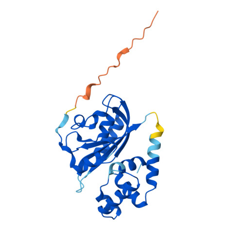 | |
UniProt | |||||
Entity Groups | |||||
| Sequence Clusters | 30% Identity50% Identity70% Identity90% Identity95% Identity100% Identity | ||||
| UniProt Group | Q2NB98 | ||||
Sequence AnnotationsExpand | |||||
Reference Sequence | |||||


