9XXU | pdb_00009xxu

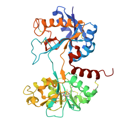
Crystal structure of the chymotrypsin-cleaved iron-free C-lobe of bovine lactoferrin at 2.82 Angstrom resolution


Experimental Data Snapshot

  • Method: X-RAY DIFFRACTION
  • Resolution: 2.82 Å
  • R-Value Free: 
    0.269 (Depositor), 0.270 (DCC) 
  • R-Value Work: 
    0.211 (Depositor), 0.216 (DCC) 

Starting Model: experimental
View more details

wwPDB Validation 3D Report Full Report

Validation slider image for 9XXU

Ligand Structure Quality Assessment 


This is version 1.1 of the entry. See complete history

Literature

Structural Basis for Single-Site Cleavage of Lactoferrin by Diverse Proteases for Prolonged Antibacterial Action: Structure of the Chymotrypsin-Cleaved Lactoferrin C-Lobe.

Pandit, S.Ahmad, N.Sharma, P.Sharma, S.Singh, T.P.

(2026) Proteins 

  • DOI: https://doi.org/10.1002/prot.70148
  • Primary Citation Related Structures: 
    9XXU

  • PubMed Abstract: 

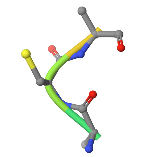
    The stable lactoferrin C-lobe offers strong potential for therapeutic applications as an antibacterial agent. Lactoferrin is a 78 kDa (Ala1Arg689) iron-binding glycoprotein which is composed of two homologous N- and C-lobes, connected by an 11-residue α-helical linker (Thr334Arg344). The limited proteolysis of lactoferrin, carried out using chymotrypsin, generated a 40 kDa, fully functional C-lobe. The structure determination revealed that the protein chain consisted of residues from Thr343 to Leu680 together with a disulfide-linked tripeptide, Ala683Cys684Ala685. It showed that the cleavage occurred specifically at the Tyr342Thr343 peptide bond within the inter-lobe 11-residue-long peptide. Remarkably, previous studies using proteinase K, trypsin, and pepsin also produced an identical C-lobe. Thus, the inter-lobe region seems to be stereochemically designed by nature for the single-site cleavage by multiple digestive enzymes. The proteolytically generated C-lobe, with three observed glycosylation sites, remains stable for 3 days in the presence of digestive enzymes. The stable C-lobe continues to sequester iron, thus showing a prolonged antibacterial property. This is a unique example of evolutionary convergence whereby multiple digestive enzymes cleave a native protein into a stable half molecule with full antibacterial action.


  • Organizational Affiliation
    • Department of Biophysics, All India Institute of Medical Sciences, New Delhi, India.

Macromolecule Content 

  • Total Structure Weight: 80.8 kDa 
  • Atom Count: 5,564 
  • Modeled Residue Count: 682 
  • Deposited Residue Count: 694 
  • Unique protein chains: 2

Macromolecules

Find similar proteins by:|  3D Structure
Entity ID: 1
MoleculeChains  Sequence LengthOrganismDetailsImage
LactotransferrinA,
C [auth B]
338Bos taurusMutation(s): 0 
EC: 3.4.21
UniProt
Find proteins for P24627 (Bos taurus)
Explore P24627 
Go to UniProtKB:  P24627
Entity Groups
Sequence Clusters30% Identity50% Identity70% Identity90% Identity95% Identity100% Identity
UniProt GroupP24627
Glycosylation
Glycosylation Sites: 3
Sequence Annotations
Expand
Reference Sequence
Find similar proteins by:  Sequence   |   3D Structure  
Entity ID: 2
MoleculeChains  Sequence LengthOrganismDetailsImage
C-terminal fragment of LactotransferrinB [auth C],
D
9Bos taurusMutation(s): 0 
EC: 3.4.21
UniProt
Find proteins for P24627 (Bos taurus)
Explore P24627 
Go to UniProtKB:  P24627
Entity Groups
UniProt GroupP24627
Sequence Annotations
Expand
Reference Sequence

Oligosaccharides

Help  
Entity ID: 3
MoleculeChains Length2D Diagram GlycosylationD Interactions
alpha-D-mannopyranose-(1-3)-[alpha-D-mannopyranose-(1-6)]beta-D-mannopyranose-(1-4)-2-acetamido-2-deoxy-beta-D-glucopyranose-(1-4)-2-acetamido-2-deoxy-beta-D-glucopyranose
E, H
5N-Glycosylation
Glycosylation Resources
GlyTouCan: G22768VO
GlyCosmos: G22768VO
GlyGen: G22768VO
Entity ID: 4
MoleculeChains Length2D Diagram GlycosylationD Interactions
alpha-D-mannopyranose-(1-2)-alpha-D-mannopyranose-(1-3)-[alpha-D-mannopyranose-(1-6)]beta-D-mannopyranose-(1-4)-2-acetamido-2-deoxy-beta-D-glucopyranose-(1-4)-2-acetamido-2-deoxy-beta-D-glucopyranose
F, G
6N-Glycosylation
Glycosylation Resources
GlyTouCan: G56014GC
GlyCosmos: G56014GC
GlyGen: G56014GC

Small Molecules

Ligands 5 Unique
IDChains Name / Formula / InChI Key2D Diagram3D Interactions
NAG

Query on NAG


Download:Ideal Coordinates CCD File
M [auth A],
Q [auth B]
2-acetamido-2-deoxy-beta-D-glucopyranose
C8 H15 N O6
OVRNDRQMDRJTHS-FMDGEEDCSA-N
SO4
(Subject of Investigation/LOI)

Query on SO4


Download:Ideal Coordinates CCD File
N [auth A],
O [auth A],
R [auth B]
SULFATE ION
O4 S
QAOWNCQODCNURD-UHFFFAOYSA-L
GOL

Query on GOL


Download:Ideal Coordinates CCD File
J [auth A],
P [auth B]
GLYCEROL
C3 H8 O3
PEDCQBHIVMGVHV-UHFFFAOYSA-N
EDO

Query on EDO


Download:Ideal Coordinates CCD File
I [auth A],
L [auth A]
1,2-ETHANEDIOL
C2 H6 O2
LYCAIKOWRPUZTN-UHFFFAOYSA-N
ACT

Query on ACT


Download:Ideal Coordinates CCD File
K [auth A]ACETATE ION
C2 H3 O2
QTBSBXVTEAMEQO-UHFFFAOYSA-M

Experimental Data & Validation

Experimental Data

  • Method: X-RAY DIFFRACTION
  • Resolution: 2.82 Å
  • R-Value Free:  0.269 (Depositor), 0.270 (DCC) 
  • R-Value Work:  0.211 (Depositor), 0.216 (DCC) 
Space Group: C 2 2 21
Unit Cell:
Length ( Å )Angle ( ˚ )
a = 92.684α = 90
b = 139.06β = 90
c = 120.741γ = 90
Software Package:
Software NamePurpose
REFMACrefinement
MxCuBEdata collection
XDSdata reduction
autoPROCdata scaling
MOLREPphasing
Cootmodel building

Structure Validation

View Full Validation Report


Ligand Structure Quality Assessment 


Entry History 

& Funding Information

Deposition Data


Funding OrganizationLocationGrant Number
Not funded--

Revision History  (Full details and data files)

  • Version 1.0: 2025-12-31
    Type: Initial release
  • Version 1.1: 2026-07-15
    Changes: Database references